Case Reports
. 2020 Jun 12;20(1):63.
doi: 10.1186/s12880-020-00463-6.

Diagnosis of foetal vein of galen aneurysmal malformation by ultrasound combined with magnetic resonance imaging: a case series

Affiliations
Case Reports

Diagnosis of foetal vein of galen aneurysmal malformation by ultrasound combined with magnetic resonance imaging: a case series

Tian-Gang Li et al. BMC Med Imaging. .

Abstract

Background: Foetal vein of Galen aneurysmal malformation (VGAM) is a very rare congenital malformation of the cerebral blood vessels. We sought to evaluate the diagnostic value of ultrasound in combination with magnetic resonance imaging (MRI) in foetal VGAM.

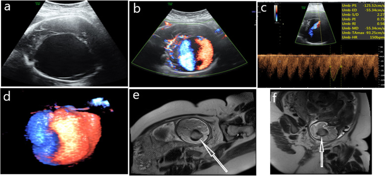
Case presentation: Prenatal ultrasound combined with MRI diagnosed five cases of VGAM. Two dimensional ultrasound images were used to find the echo-free cystic structure below the thalamus and above the cerebellum with five cases. Colour blood flow showed dilated VGAM in five cases, while the arteriovenous spectrum was explored in two cases and foetal heart failure was found in other three cases. MRI was manifested as a dilated VGAM found at the midline of the brain, demonstrating widening or dilation of the straight sinus in four cases, ventricular dilatation in one case, brain parenchyma bleeding in two cases, and grey matter softening in one case. One infant died on the day of its birth, while the other four infants died within one month to six months after birth.

Conclusions: Ultrasound combined with MRI can more accurately and comprehensively observe the pathological characteristics of VGAM, diagnose related complications early and determine its prognosis.

Keywords: Congenital malformation; Foetus, vein of Galen aneurysmal malformation; Prenatal diagnosis; Ultrasound, magnetic resonance imaging.


Conflict of interest statement

The authors declare that they have no competing interests.

Figures

Fig. 1
Fig. 1
Case 1, 35-week prenatal ultrasound and MRI images of a VGAM foetus. a: Two-dimensional ultrasound shows a huge thin-walled echoless area next to the midline of the brain. b: Colour Doppler shows a full blood-flow signal in the brain’s echoless area. c: The skull’s echoless area shows a high-speed low-impedance blood-flow spectrum. d: The reconstructed three-dimensional energy Doppler image clearly shows the dilated Galen vein, but no obvious straight sinus is displayed. e: MRI cross-section of the foetal skull showing the dilated Galen vein through flow phenomenon (white arrow). f: Sagittal section of the foetal brain showing dilated Galen’s venous aneurysm by flow phenomenon (white arrow)
Fig. 2
Fig. 2
Case 2, 30-week prenatal ultrasound and MRI images of a VGAM foetus. a: Two-dimensional ultrasound shows a huge thin-walled echoless area next to the midline of the brain. b: Colour Doppler shows a full blood-flow signal in the brain’s echoless area. c: The skull’s echoless area shows arteriovenous leak with arteriovenous mixed spectrum; d: The reconstructed three-dimensional energy Doppler image clearly shows the dilated VGAM, and obvious straight sinus is displayed (white arrow). e: MRI cross-section of the foetal skull showing the dilated VGAM through flow phenomenon (shown by white arrow). f: Sagittal section of the foetal brain showing the dilated Galen’s venous aneurysm by flow phenomenon (white arrow)

Similar articles

Cited by

References

    1. Smith A, Abruzzo T, Mahmoud M. Vein of Galen malformation and high-output cardiac failure. Anesthesiology. 2016;125(3):597. doi: 10.1097/ALN.0000000000001095. - DOI - PubMed
    1. Mortazavi MM, Griessenauer CJ, Foreman P, Bavarsad Shahripour R, Shoja MM, Rozzelle CJ, et al. Vein of Galen aneurysmal malformations: critical analysis of the literature with proposal of a new classification. J Neurosurg Pediatr. 2013;12(3):293–306. doi: 10.3171/2013.5.PEDS12587. - DOI - PubMed
    1. Deloison B, Chalouhi GE, Sonigo P, Zerah M, Millischer AE, Dumez Y, et al. Hidden mortality of prenatally diagnosed vein of Galen aneurysmal malformation: retrospective study and review of the literature. Ultrasound Obstet Gynecol. 2012;40(6):652–658. doi: 10.1002/uog.11188. - DOI - PubMed
    1. Sepulveda W, Vanderheyden T, Pather J, Pasquini L. Vein of galen malformation:prenatal evaluation with three-dimensional power Doppler angiography. J Ultrasound Med. 2003;22(12):1395–1398. doi: 10.7863/jum.2003.22.12.1395. - DOI - PubMed
    1. Félix L, Souza AR, Queiroz AP, Diniz C, Lima M, Santos RE, et al. Prenatal ultrasonography in the diagnosis of vein of Galen aneurysm. Acta Medica Port. 2010;23(3):505–510. - PubMed

Publication types

Supplementary concepts