Review
. 2020 Oct;18(10):571-586.
doi: 10.1038/s41579-020-0385-0. Epub 2020 Jun 12.

Biofilm dispersion

Affiliations
Review

Biofilm dispersion

Kendra P Rumbaugh et al. Nat Rev Microbiol. 2020 Oct.

Abstract

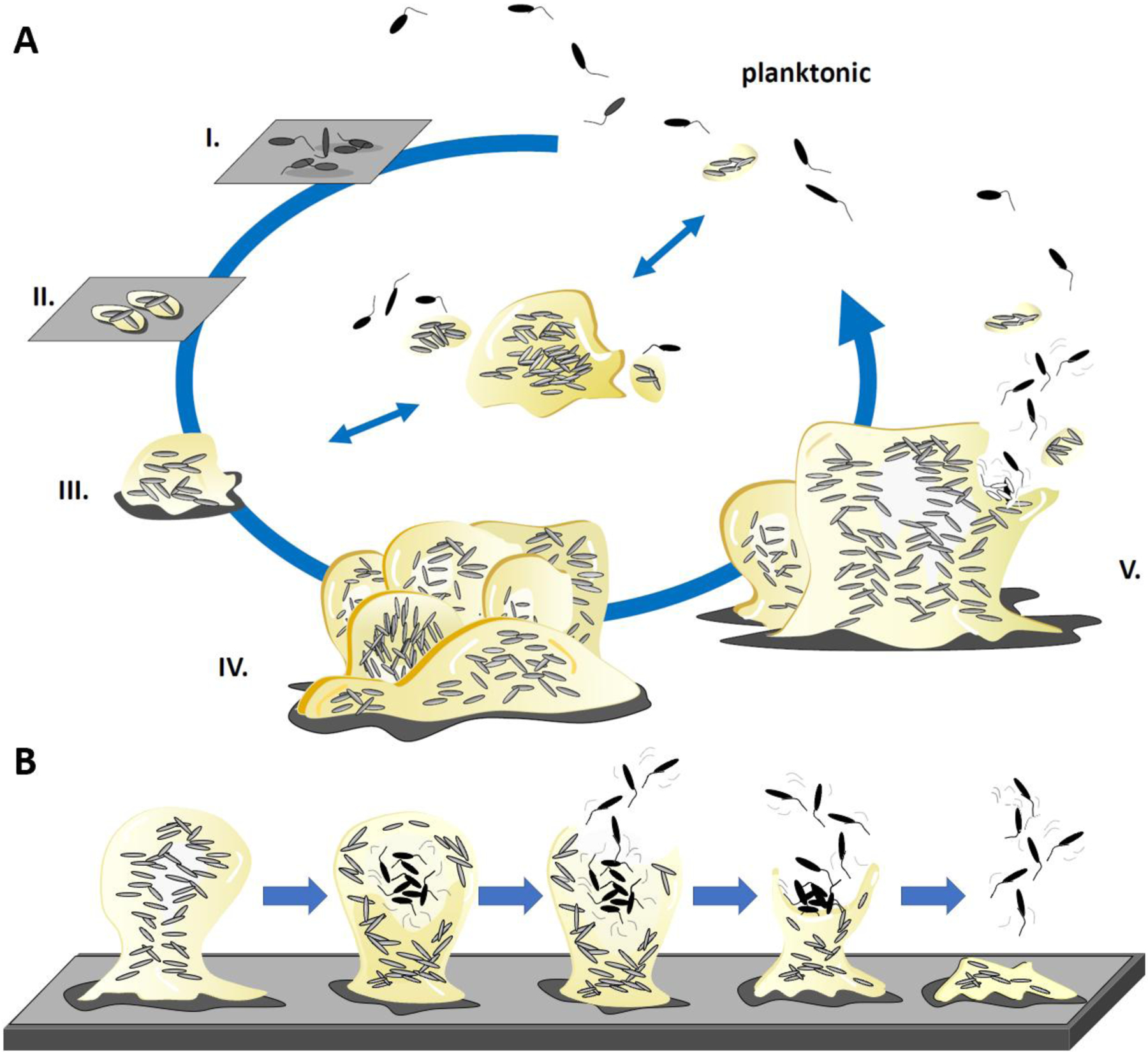
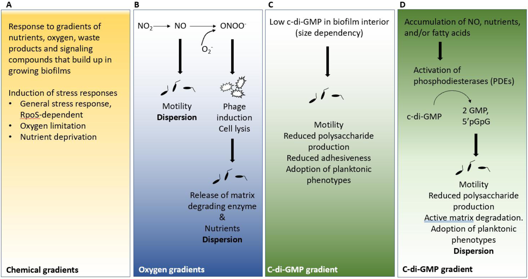
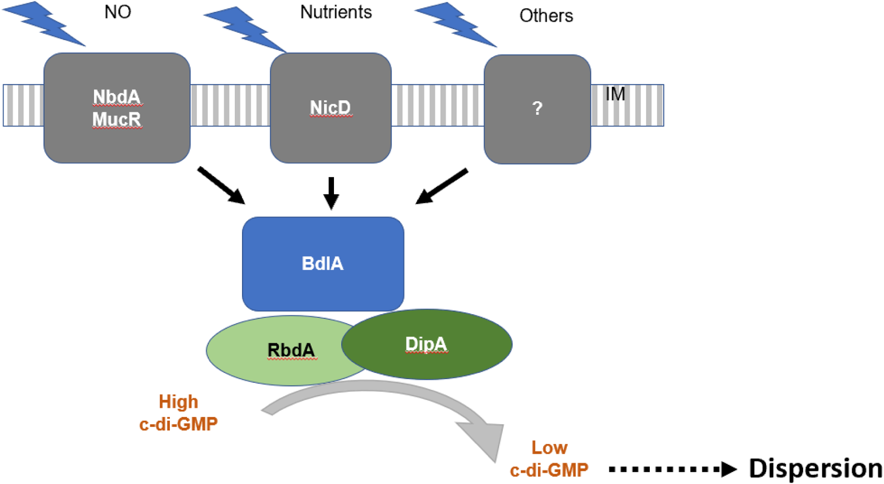
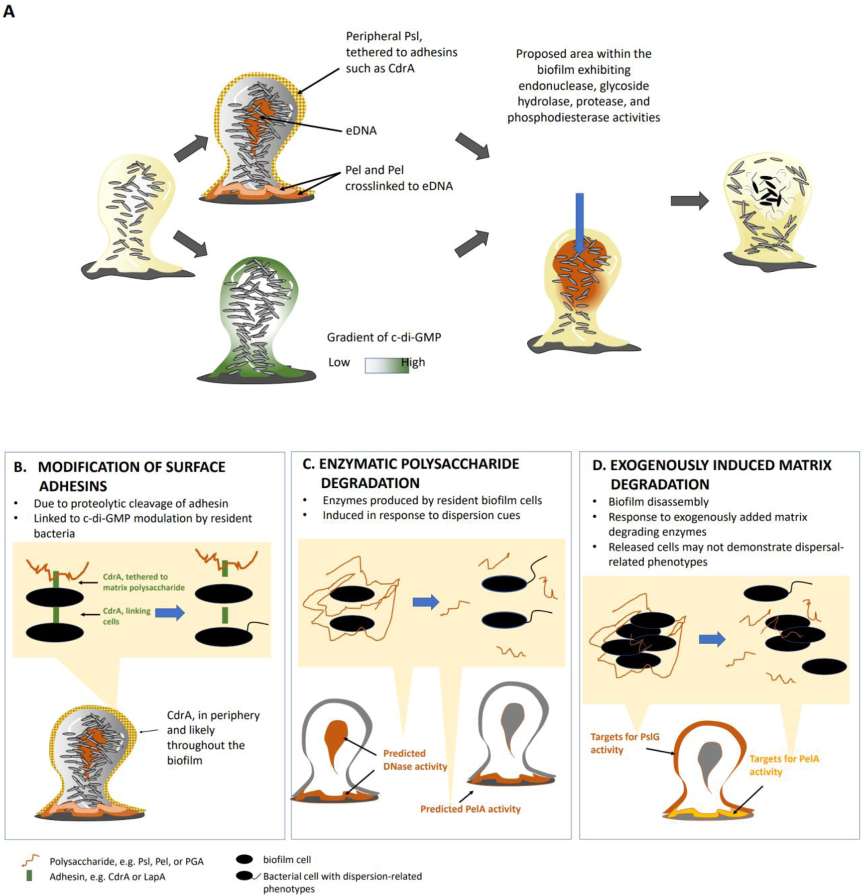
The formation of microbial biofilms enables single planktonic cells to assume a multicellular mode of growth. During dispersion, the final step of the biofilm life cycle, single cells egress from the biofilm to resume a planktonic lifestyle. As the planktonic state is considered to be more vulnerable to antimicrobial agents and immune responses, dispersion is being considered a promising avenue for biofilm control. In this Review, we discuss conditions that lead to dispersion and the mechanisms by which native and environmental cues contribute to dispersion. We also explore recent findings on the role of matrix degradation in the dispersion process, and the distinct phenotype of dispersed cells. Last, we discuss the translational and therapeutic potential of dispersing bacteria during infection.


Conflict of interest statement

Competing interests

The authors declare no competing interests.

Figures

Figure 1:
Figure 1:. Biofilm formation and dispersion.
a) The schematic is based on the in vitro analysis of single species Pseudomonas aeruginosa biofilms. The formation of biofilms is a cyclic process that occurs in a stage-specific and progressive manner. The process is initiated following surface contact by single planktonic cells. Several developmental steps are discernable as reversible attachment (step I), irreversible attachment (step II) and biofilm maturation (steps III-IV),. During the initial event in biofilm development, bacteria attach to substratum at the cell pole or via the flagellum (step I). Cells are cemented to the substratum and form nascent cell clusters with all cells in contact with the substratum. Transition to this stage coincides with loss of flagella gene expression and the production of biofilm matrix components (step II). Cell clusters mature and are several cells thick, embedded in the extracellular polymeric substances matrix (step III). Biofilms fully mature, which is apparent by clusters and microcolonies having reached maximum thickness (step IV). The biofilm life cycle comes full circle when biofilms disperse (step V). This stage is characterized by cells evacuating the interior portions of cell clusters, forming void spaces. Recent reports describe the formation of aggregates that exhibit biofilm-like features in the absence of surfaces (middle panel). Based on in vivo observations, these aggregates can attach to surfaces or disperse (indicated by the arrows). However, it is unclear if aggregate formation requires the same biofilm formation pathways and dispersion events. b) The schematic shows the discernable events that lead to biofilm dispersion. During dispersion, the inside of a biofilm becomes fluid, and cells within this zone begin to show signs of agitation and movement. Subsequently, cells escape the biofilm via a disruption in the microcolony wall through which cells evacuate, entering the bulk liquid as single bacteria. This leaves behind biofilms with central voids. If the dispersion response is extensive, the biofilm structure may further erode.
Figure 2:
Figure 2:. Environmental conditions initiating dispersion.
Each panel indicates various chemical gradients that are likely to be present in biofilms; low concentrations of those chemicals have been linked to dispersion. Moreover, phenotypic changes such as motility or induction of events that are triggered in response to the limited availability of compounds are shown. a) Bacteria residing at different locations within the biofilm structure experience concentration gradients of nutrient resources, oxygen, waste products and extracellular signaling molecules. Biofilm cells respond to these gradients by inducing various stress responses, such as the RpoS -dependent general stress response, as well as increased expression of genes involved in the response to oxygen limitation and nutrient deprivation. b) Under limiting oxygen conditions, such as found in the interior of the biofilm, anaerobic denitrification leads to the generation of nitric oxide (NO). Exposure to NO is linked to the reduction in cellular bis-(3’-5’)-cyclic dimeric guanosine monophosphate (c-di-GMP) levels and thus increased motility and dispersion. Furthermore, NO can react with superoxide to generate the cell-toxic radical ONOO. ONOO causes cellular damage, bacteriophage induction and cell lysis. Cell lysis results in the release of degradative enzymes that contribute to the enzyme-mediated breakdown of the biofilm matrix and/or loosening of the biofilm matrix. In addition, cell lysis generates nutrients for growth. c)-d) Levels of c-di-GMP vary throughout the biofilm, with the lowest levels being detected in the biofilm interior. Low c-di-GMP levels contribute to phenotypes generally associated with the planktonic mode of growth, including increased motility, increased drug susceptibility, but reduced adhesiveness and matrix production. Bacteria residing at different locations within the biofilm structure experience the accumulation of native dispersion cues including NO, cis-DA, and nutrients. Sensing of dispersion cues activates phosphodiesterases (PDEs) capable of degrading c-di-GMP, ultimately resulting in an overall reduction in the levels of c-di-GMP. Phenotypes associated with low c-di-GMP levels include increased motility, reduced adhesiveness, reduced matrix production and dispersion.
Figure 3:
Figure 3:. Sensing of dispersion cues.
Model of dispersion cue sensing and modulation of bis-(3’-5’)-cyclic dimeric guanosine monophosphate (c-di-GMP) levels to induce dispersion. In Pseudomonas aeruginosa, dispersion cue sensing involves the chemotaxis-like MCP homolog, BdlA, the c-di-GMP phosphodiesterases (PDEs) DipA, RbdA, MucR and NbdA, and the diguanylate cyclases (DGCs) GcbA and NicD. The membrane-associated PDEs MucR and NbdA and the DGC NicD are involved in perceiving and relaying dispersion cues to promote the modulation of c-di-GMP levels. The DGC NicD and the PDE NbdA contribute to dispersion in a cue-specific manner, with NbdA sensing NO and NicD sensing nutrient cues. Receptors for other dispersion cues such as fatty acids have not yet been elucidated (indicated by the question mark). BdlA and the PDEs DipA and RbdA are central to the dispersion response regardless of the cue that is being sensed and contribute to the overall reduction of c-di-GMP levels post dispersion cue sensing.
Figure 4.
Figure 4.. Mechanisms resulting in biofilm dispersal.
(a) Model of spatial localization of biofilm matrix components and bis-(3’-5’)-cyclic dimeric guanosine monophosphate (c-di-GMP), and proposed biofilm areas with active matrix- and c-di-GMP-degrading activities, that lead to biofilm dispersion. In Pseudomonas aeruginosa PAO1, the matrix polysaccharide Psl is localized at the periphery and the base of biofilms. Psl interacts with the adhesin protein CdrA and forms a protective but non-rigid structure in between cells and around the biofilm structure. The Pel polysaccharide is primarily located at the base of the biofilm and crosslinked to extracellular DNA (eDNA). However, eDNA is not limited to the biofilm base but has also been detected in the biofilm interior. Mature biofilms are characterized by low c-di-GMP levels in the interior of the biofilm whereas c-di-GMP levels in immature or less structured biofilms is more uniform. Considering the link between dispersion, low c-di-GMP levels, and matrix degradation, the biofilm periphery and biofilm interior are the most likely to be the locations within the biofilm that experiecne increased matrix- and c-di-GMP-degrading activities. Moreover, these locations coincide with observed dispersion events such as void formation and erosion of the biofilm structure. b) Surface adhesins such as CdrA or LapA are cleaved to untether the polysaccharide matrix and break cell-cell interactions. (c) Matrix components are enzymatically degraded (by intrinsic DNases or the hydrolase PelA) in response to native or environmental dispersion cues (nitric oxide, nutrients or cis-2-decenoic acid). d) Biofilm disassembly can be induced by exogenously added matrix-degrading enzymes. As enzymes are exogenously added, matrix degradation is expected to primarily target the periphery of the biofilm structure (see Box 1).
Figure 5.
Figure 5.. Bacterial dissemination triggered by glycoside hydrolase treatment of Pseudomonas aeruginosa biofilms.
In vitro image system (IVIS) imaging shows that treatment of 48-hour-old mouse chronic wounds, infected with bioluminescent P. aeruginosa, with α-amylase and cellulase (glycoside hydrolase treatment), resulted in systemic spread of the infection (bottom panles). By contrast, systemic spread was not obeserved after treatment with heat-inactivated enzymes (top panels). Modified from Ref..

References

    1. Costerton JW, Stewart PS & Greenberg EP Bacterial biofilms: a common cause of persistent infections. Science 284, 1318–1322 (1999). - PubMed
    1. Flemming H-C et al. Biofilms: an emergent form of bacterial life. Nature Reviews Microbiology 14, 563, doi:10.1038/nrmicro.2016.94 (2016). - DOI - PubMed
    1. Geesey GG, Richardson WT, Yeomans HG, Irvin RT & Costerton JW Microscopic examination of natural sessile bacterial populations from an alpine stream. Can. J. Microbiol 23, 1733–1736 (1977). - PubMed
    1. Costerton JW et al. Bacterial biofilms in nature and disease. Annual Reviews in Microbiology 41, 435–464 (1987). - PubMed
    1. Stoodley P, Sauer K, Davies DG & Costerton JW Biofilms as complex differentiated communities. Annual Reviews in Microbiology 56, 187–209, doi:doi:10.1146/annurev.micro.56.012302.160705 (2002). - DOI - PubMed

Publication types

MeSH terms