. 2020 Aug;34(8):10267-10285.
doi: 10.1096/fj.202000629R. Epub 2020 Jun 13.

Resident mesenchymal vascular progenitors modulate adaptive angiogenesis and pulmonary remodeling via regulation of canonical Wnt signaling

Affiliations

Resident mesenchymal vascular progenitors modulate adaptive angiogenesis and pulmonary remodeling via regulation of canonical Wnt signaling

Megan E Summers et al. FASEB J. 2020 Aug.

Abstract

Adaptive angiogenesis is necessary for tissue repair, however, it may also be associated with the exacerbation of injury and development of chronic disease. In these studies, we demonstrate that lung mesenchymal vascular progenitor cells (MVPC) modulate adaptive angiogenesis via lineage trace, depletion of MVPC, and modulation of β-catenin expression. Single cell sequencing confirmed MVPC as multipotential vascular progenitors, thus, genetic depletion resulted in alveolar simplification with reduced adaptive angiogenesis. Following vascular endothelial injury, Wnt activation in MVPC was sufficient to elicit an emphysema-like phenotype characterized by increased MLI, fibrosis, and MVPC driven adaptive angiogenesis. Lastly, activation of Wnt/β-catenin signaling skewed the profile of human and murine MVPC toward an adaptive phenotype. These data suggest that lung MVPC drive angiogenesis in response to injury and regulate the microvascular niche as well as subsequent distal lung tissue architecture via Wnt signaling.

Keywords: Wnt signaling; adaptive angiogenesis; emphysema; mesenchymal vascular progenitor cell; microvascular niche.


Conflict of interest statement

The authors have no conflicts of interest to declare.

Figures

FIGURE 1
FIGURE 1
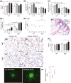
Isolated Abcg2pos MVPC have angiogenic transcriptional signatures and potential. A‐C, Abcg2‐Cre‐ERT2 Rosa26mTmG WT mice were induced with intraperitoneal tamoxifen. Two days post induction mice were sacrificed, and lungs agarose inflated using constant pressure, to obtain lung tissue for precision cut lung slices for two‐photon imaging. Membrane labeled eGFP MVPC were visible in green and mTomato lung tissue was detected in the red channel. A, Representative 2 µM section through the lung tissue Z stack. B and C, Reconstruction of the three‐dimensional lung image naïve and with a Gaussian filter. Scale and grid dimension = 20 µM. D, Abcg2‐Cre‐ERT2 Rosa26mTmG WT mice were induced with intraperitoneal tamoxifen. Two days post induction mice were sacrificed, and lung tissue digested to a single cell suspension for cell sorting to obtain the eGFP labeled cells. E, t‐SNE plot depicting CD45neg eGFP labeled cells analyzed using 10x single cell RNA sequencing. F, GO clustering analysis. G and H, Angiogenic sprouting and migration potential of MVPC was defined by co‐culture three‐dimensional spheroid assays
FIGURE 2
FIGURE 2
Targeted depletion of MVPC effect on distal lung structure and adaptive angiogenesis. WT or f/fSTOP DTA Abcg2‐Cre‐ERT2 Rosa26mTmG mice were induced with intraperitoneal tamoxifen. One month or 15 months following induction mice were sacrificed, and lungs agarose inflated using constant pressure, to obtain lung tissue for histological and immunofluorescent analyses. n = 4,5 (1 month). A, Quantitation of MLI. B, Fractional volume, the fraction of an image that is occupied by lung tissue. C and D, Representative H&E stained lung tissue sections. Scale bar = 50 µM. n = 10, 12 (15 months) E and F, Representative H&E stained lung tissue sections. Scale bar = 100 µM. G. Quantitation of MLI. H and I, Mean compliance and resistance measured by FlexiVent. WT, f/fSTOP DTA mice were induced with intraperitoneal tamoxifen, 2 weeks later mice were exposed to cigarette smoke for four weeks. Six weeks following induction mice were sacrificed, and lungs agarose inflated using constant pressure, to obtain lung tissue for histological analyses. n = 4, 9, 4, 5. K, Quantitation of MLI and L, surface to volume ratio. Immunostaining was performed on lung tissue sections to detect smooth muscle alpha actin (SMA) and F8 positive microvessels as well as muscularization. M‐O, The immune‐positive microvessels were counted per field of view. A 6‐8 sections of 20 field of view (f.o.v.) per section were evaluated
FIGURE 3
FIGURE 3
Canonical Wnt activation in Abcg2pos MVPC blunted adaptive responses to hypoxia and exacerbated injury following Sugen‐hypoxia exposure. Adult WT or βcatenin over expresser (βOE) mice were treated with intraperitoneal tamoxifen. Two weeks following induction mice were grouped for exposure to room air (RA) + CMC vehicle (Veh.), room air + Sugen5416 (SU5416), hypoxia + CMC vehicle (10% oxygen; HY Veh.) or hypoxia + SU5416 (HY SU). Histological and physiological parameters were analyzed after three weeks. A, A pressure transducer was placed in the jugular to the right heart to measure RVSP. Lungs were agarose inflated using constant pressure to obtain lung tissue for histological and immunofluorescent analyses. Immunostaining was performed on lung tissue sections to detect (B) smooth muscle alpha actin (SMA) and (C) F8 positive microvessels as well as muscularization. The immune‐positive microvessels were counted per field of view. A 6‐8 sections of 20 field of view (f.o.v.) per section were evaluated. D, Trichrome staining was performed on tissue sections and collagen positive microvessels were enumerated. A 6‐8 sections of 20 f.o.v. per section were examined. E, Total collagen per f.o.v. was also calculated using FIJI software. F, Representative image of fibrosis in a trichrome stained βOE hypoxia + SU5416 lung section. G‐J, Representative trichrome images illustrate distal lung tissue structure. Scale bars = 100 µM. K, MLI was calculated to evaluate simplification of distal lung tissue structure. Data presented as the mean (±SEM). n = 6‐9 mice/group. RA Vehicle (white box); RA SU5416 (light grey box); HY Vehicle (dark grey box); HY SU5416 (black box).L‐N, Angiogenic sprouting and migration potential of primary eGFPpos MVPC was defined by co‐culture three‐dimensional spheroid assays (n = 10, 10)
FIGURE 4
FIGURE 4
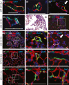
MVPC directly participate in de novo angiogenesis following lung injury. Immunostaining was performed on lung tissue sections from room air (RA) + CMC vehicle (Veh.), room air + Sugen5416 (SU5416), hypoxia (10% oxygen) or hypoxia + SU groups to detect eGFP labeled MVPC and lineage derivatives. Costaining was performed to detect SMA positive muscularized vessels, Factor VIII (F8; G&I) or podoplanin (podopln; J‐O) positive microvascular endothelium. Representative images from WT or βOE hypoxia + SU groups were presented. A, MVPC migrate along vascular wall. B‐D, MVPC form vascular structures via bridging or sprouting adjacent to SMApos microvessels. E‐I, Fibrosis was identified in the βOE hypoxia + SU groups. E, Trichrome staining. F, Immunofluorescent detection of eGFP and SMA. G, Enlarged from F, Immunofluorescent detection of eGFP, SMA, and FactorVIII. H, Enlarged eGFP expressing vascular structure from G. I. Enlarged eGFP F8 negative vascular structure from G, H. J, Immunofluorescent detection of eGFP and podoplanin in WT lungs. K, Enlarged eGFP expressing structures adjacent to podoplanin positive endothelium from J. L, Migrating MVPC do not express podoplanin. M, Immunofluorescent detection of eGFP and podoplanin in βOE lungs enlarged in N and O. N, eGFP adjacent to podoplanin positive endothelium. O, eGFP positive structures lacking podoplanin expressing endothelium. Nuclei are stained with DAPI (blue). The presence of nuclei in MVPC derived structures was indicated by the presence of *. n = 6‐9 mice/group. Scale bars = 100 µM
FIGURE 5
FIGURE 5
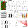
Disrupted Wnt signaling in ABCG2pos COPD MVPC disrupts MVEC function. A, Heatmap for differentially expressed genes in MVPC from COPD vs non‐diseased lungs (color scale shown at the top right) B and C, Validation of representative genes expressed in COPD ABCG2pos lung MVPC, compared to control, enriched in GO categories of B, Wnt signaling and C, actin binding, contractility, and migration. The mean of combined patient samples per group as well as results for individual samples are presented. n = 3‐6 patient samples per group. D, Western blot analysis of Dkk1, Wnt5a and Slit2 protein expression. n = 3, 3. Data presented as the mean ± standard error of mean (SEM). E, Cocultures of pulmonary MVEC and MVPC were analyzed for the effect of control or COPD MVPC on barrier function following injury. MVPC were plated on a monolayer of MVEC at a ratio of 3:1. The groups underwent no injury or an electrical wounding injury using the ECIS system. The presence of COPD MVPC following injury decreased the rate at which barrier formation is recovered. Quantitation of normalized resistance at indicated time points (Δ) was presented in bar graph format. Data presented as mean (±SEM). Controls included MVEC alone and uninjured MVEC. n = 4. F, Murine WT and Wnt activated βOE MVPC were analyzed by PCR to examine the expression of angiogenic transcripts identified as different between control and COPD samples. n = 3, 3. All amplification was normalized to a housekeeping gene and the results presented as mean fold change over control. Data presented as mean ± SEM. G. Representative summary of murine and human PCR data
FIGURE 6
FIGURE 6
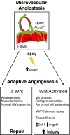
Wnt signaling in the Abcg2HI MVPC regulate adaptive responses of the microvasculature. Wnt signaling and function of MVPC is regulated by the disease microenvironment, to influence the microvascular program of adaptation resulting in either repair or remodeling. The microvascular program of adaptation includes: angiogenesis vs microvessel rarefaction, muscularization of microvessels or collagen deposition and the MVPC direct contribution to the formation of angiogenic tubes. MVPC depicted in green and microvascular endothelial cells (MVEC) in red

References

    1. Sabit R, Bolton CE, Edwards PH, et al. Arterial stiffness and osteoporosis in chronic obstructive pulmonary disease. Am J Respir Crit Care Med. 2007;175:1259‐1265. - PubMed
    1. McAllister DA, Maclay JD, Mills NL, et al. Arterial stiffness is independently associated with emphysema severity in patients with chronic obstructive pulmonary disease. Am J Respir Crit Care Med. 2007;176:1208‐1214. - PMC - PubMed
    1. Barr RG, Mesia‐Vela S, Austin JHM, et al. Impaired flow‐mediated dilation is associated with low pulmonary function and emphysema in ex‐smokers: the Emphysema and Cancer Action Project (EMCAP) Study. Am J Respir Crit Care Med. 2007;176:1200‐1207. - PMC - PubMed
    1. Yu N, Wei X, Li Y, Deng L, Jin C‐W, Guo Y. Computed tomography quantification of pulmonary vessels in chronic obstructive pulmonary disease as identified by 3D automated approach. Medicine. 2016;95:e5095. - PMC - PubMed
    1. Alford SK, van Beek EJR, McLennan G, Hoffman EA. Heterogeneity of pulmonary perfusion as a mechanistic image‐based phenotype in emphysema susceptible smokers. Proc Natl Acad Sci U S A. 2010;107:7485‐7490. - PMC - PubMed

Publication types

MeSH terms