

Targeted manipulation of pain neural networks: The potential of focused ultrasound for treatment of chronic pain
- PMID: 32534900
- PMCID: PMC7483565
- DOI: 10.1016/j.neubiorev.2020.06.007
Targeted manipulation of pain neural networks: The potential of focused ultrasound for treatment of chronic pain
Abstract
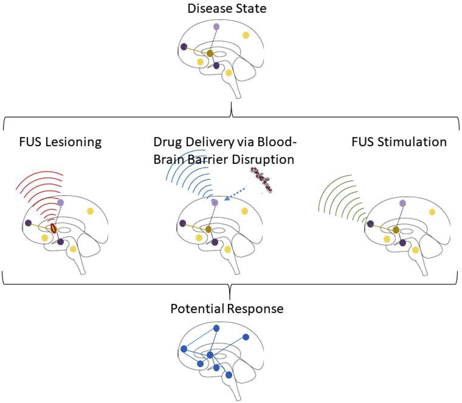
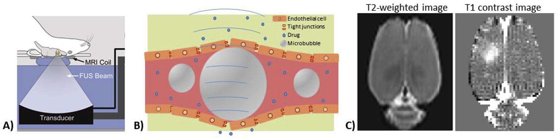
Focused ultrasound (FUS) is a promising technology for facilitating treatment of brain diseases including chronic pain. Focused ultrasound is a unique modality for delivering therapeutic levels of energy into the body, including the central nervous system (CNS). It is non-invasive and can target spatially localized effects through the intact skull to cortical or subcortical regions of the brain. FUS can achieve three different mechanisms of action in the brain that are relevant for chronic pain treatment: (1) localized thermal ablation of neural tissue; (2) localized and transient disruption of the blood-brain barrier for targeted drug delivery to CNS structures; and (3) inhibition or stimulation of neuronal activity in targeted regions. This review provides an in-depth look at the technology of FUS with emphasis placed on applications to CNS-based treatments of chronic pain. While still in the early stages of clinical translation and with some technical challenges remaining, we suggest that FUS has great potential as a novel approach for manipulating CNS networks involved in pain treatment.
Keywords: Analgesia; Brain; Focused ultrasound; Neural circuits; Neuromodulation; Neurosurgical ablation; Pain; Targeted drug delivery.
Copyright © 2020 Elsevier Ltd. All rights reserved.
Figures
Similar articles
-
Short-Term Memory Impairment.2024 Jun 8. In: StatPearls [Internet]. Treasure Island (FL): StatPearls Publishing; 2025 Jan–. 2024 Jun 8. In: StatPearls [Internet]. Treasure Island (FL): StatPearls Publishing; 2025 Jan–. PMID: 31424720 Free Books & Documents.
-
The Black Book of Psychotropic Dosing and Monitoring.Psychopharmacol Bull. 2024 Jul 8;54(3):8-59. Psychopharmacol Bull. 2024. PMID: 38993656 Free PMC article. Review.
-
Secondary effects on brain physiology caused by focused ultrasound-mediated disruption of the blood-brain barrier.J Control Release. 2020 Aug 10;324:450-459. doi: 10.1016/j.jconrel.2020.05.040. Epub 2020 May 26. J Control Release. 2020. PMID: 32470359 Free PMC article. Review.
-
Non-invasive brain stimulation techniques for chronic pain.Cochrane Database Syst Rev. 2018 Apr 13;4(4):CD008208. doi: 10.1002/14651858.CD008208.pub5. Cochrane Database Syst Rev. 2018. PMID: 29652088 Free PMC article.
-
Non-invasive brain stimulation techniques for chronic pain.Cochrane Database Syst Rev. 2018 Mar 16;3(3):CD008208. doi: 10.1002/14651858.CD008208.pub4. Cochrane Database Syst Rev. 2018. Update in: Cochrane Database Syst Rev. 2018 Apr 13;4:CD008208. doi: 10.1002/14651858.CD008208.pub5. PMID: 29547226 Free PMC article. Updated.
Cited by
-
Network targets for therapeutic brain stimulation: towards personalized therapy for pain.Front Pain Res (Lausanne). 2023 Jun 8;4:1156108. doi: 10.3389/fpain.2023.1156108. eCollection 2023. Front Pain Res (Lausanne). 2023. PMID: 37363755 Free PMC article.
-
Anatomo-physiological basis and applied techniques of electrical neuromodulation in chronic pain.J Anesth Analg Crit Care. 2024 May 2;4(1):29. doi: 10.1186/s44158-024-00167-1. J Anesth Analg Crit Care. 2024. PMID: 38698460 Free PMC article. Review.
-
Low frequency ultrasound elicits broad cortical responses inhibited by ketamine in mice.Commun Eng. 2024 Aug 27;3(1):120. doi: 10.1038/s44172-024-00269-2. Commun Eng. 2024. PMID: 39192002 Free PMC article.
-
Pain-preferential thalamocortical neural dynamics across species.Nat Hum Behav. 2024 Jan;8(1):149-163. doi: 10.1038/s41562-023-01714-6. Epub 2023 Oct 9. Nat Hum Behav. 2024. PMID: 37813996
-
Transient ultrasound stimulation has lasting effects on neuronal excitability.Brain Stimul. 2021 Mar-Apr;14(2):217-225. doi: 10.1016/j.brs.2021.01.003. Epub 2021 Jan 12. Brain Stimul. 2021. PMID: 33444809 Free PMC article.
References
Publication types
MeSH terms
Grants and funding
LinkOut - more resources
Full Text Sources
Medical
