Multicenter Study
. 2020 Jun:56:102804.
doi: 10.1016/j.ebiom.2020.102804. Epub 2020 Jun 11.

DRH1 - a novel blood-based HPV tumour marker

Affiliations
Multicenter Study

DRH1 - a novel blood-based HPV tumour marker

Thomas Weiland et al. EBioMedicine. 2020 Jun.

Abstract

Background: To date, no studies have successfully shown that a highly specific, blood-based tumour marker to detect clinically relevant HPV-induced disease could be used for screening, monitoring therapy response or early detection of recurrence. This study aims to assess the clinical performance of a newly developed HPV16-L1 DRH1 epitope-specific serological assay.

Methods: In a multi-centre study sera of 1486 patients (301 Head and Neck Squamous Cell Carcinoma (HNSCC) patients, 12 HIV+ anal cancer patients, 80 HIV-positive patients, 29 Gardasil-9-vaccinees, 1064 healthy controls) were tested for human HPV16-L1 DRH1 antibodies. Analytical specificity was determined using WHO reference-sera for HPV16/18 and 29 pre- and post-immune sera of Gardasil-9-vaccinees. Tumour-tissue was immunochemically stained for HPV-L1-capsidprotein-expression.

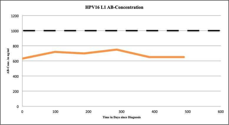
Findings: The DRH1-competitive-serological-assay showed a sensitivity of 95% (95% CI, 77.2-99.9%) for HPV16-driven HNSCC, and 90% (95% CI, 55.5-99.7%) for HPV16-induced anal cancer in HIV-positives. Overall diagnostic specificity was 99.46% for men and 99.29% for women ≥ 30 years. After vaccination, antibody level increased from average 364 ng/ml to 37,500 ng/ml. During post-therapy-monitoring, HNSCC patients showing an antibody decrease in the range of 30-100% lived disease free over a period of up to 26 months. The increase of antibodies from 2750 to 12,000 ng/ml mirrored recurrent disease. We can also show that the L1-capsidprotein is expressed in HPV16-DNA positive tumour-tissue.

Interpretation: HPV16-L1 DRH1 epitope-specific antibodies are linked to HPV16-induced malignant disease. As post-treatment biomarker, the assay allows independent post-therapy monitoring as well as early diagnosis of tumour recurrence. An AUC of 0.96 indicates high sensitivity and specificity for early detection of HPV16-induced disease.

Funding: The manufacturer provided assays free of charge.

Keywords: Antibodies; Blood test; HNSCC; HPV16; Screening; Tumour marker.


Conflict of interest statement

Declaration of Competing Interest All participating authors hereby disclose any financial and personal relationships with other people or organisations that could have inappropriately influenced the current study.

Figures

Fig 1
Fig. 1
Remark Diagram of study population and patient subsets used for calculation of sensitivity, specificity, positive predictive value, negative predictive value, ROC- and Area under the curve calculation.
Fig 2
Fig. 2
a–d. Characteristic antibody graphs during follow up. Fig. 2a: Classical antibody decrease during follow up, indicating a successful treatment understood as successful removal of tumour cells, which is associated with disease free overall surveillance. The black spotted bar at 1000 ng/ml represents the cut off antibody concentration discriminating positive and negative DRH1 test results. Patient characteristics of patient 10 as described in Table 1: Male, 58 year old, carcinoma of the tonsils, HPV16 DNA positive, p16 positive Therapy: Surgery + adjuvant Radiotherapy Decrease of antibody concentration by 90% within 6 months, and 100% within 18 months.
Fig 2
Fig. 2
a–d. Characteristic antibody graphs during follow up. Fig. 2b: Antibody concentration during follow up of patient No 12. Initial antibody decrease during follow up by 43% after 9 months (in green), indicating a successful treatment, was followed by a sudden increase of antibody concentration (in red) 3 months later– soon after tumour recurrence in the lungs was diagnosed. The black spotted bar at 1000 ng/ml represents the cut off antibody concentration discriminating positive and negative DRH1 test results. Patient characteristics of patient 12 as described in Table 1: Male, 81 year old, base of tongue carcinoma, HPV16 DNA positive, p16 positive Therapy: Radioimmunotherapie with Cetuximab *Time of diagnosis of tumour recurrence
Fig 2
Fig. 2
a–d. Characteristic antibody graphs during follow up. Fig. 2c: Characteristical graph in a patient with an HPV33 associated OPSCC showing HPV16 L1 antibody concentration at a constant low level indicating the type-specificity of the assay. The black spotted bar at 1000 ng/ml represents the cut off antibody concentration discriminating positive and negative DRH1 test results. Patient characteristics of patient 9 as described in Table 1: Male, 72 years old, base of tongue carcinoma, HPV33 DNA positive, p16 positive Therapy: Radiochemotherapy
Fig 2
Fig. 2
a–d. Characteristic antibody graphs during follow up. Fig. 2d: Overview of antibody concentrations of all HNSCC patients from Graz during follow up, baseline characteristics can be seen in Table 1. Green curves: HPV16-L1 immunoassay positive, Orange curves: HPV16-L1 immunoassay negative, Red curves: Increasing antibody titers in three HPV16-L1 immunoassay positive patients.
Fig 3
Fig. 3
Receiver-Operating-Characteristic (ROC)–curve analysis, with an ´area under the curve´ of 0.96 (95% confidence interval 0.91–1), was calculated for 20 HPV16 driven OPSCCs and 1064 controls.
Fig 4
Fig. 4
: HPV16-L1 capsid protein expression in tumour cells As shown in pictures a - d, about half of the tumour cells show a nuclear staining in red colour for the L1 capsid protein. L1 expression was confirmed by Western Blot analysis. Magnification: 100x (a), 200x (b, c), 400x (d) Pictures e and f show an `inner border like´ staining. This means different clusters of tumour cells next to each other being L1 capsid protein positive (on the right) and L1 negative (on the left). Picture f shows a higher magnification of the area marked by a white circle. Magnification: 400x (e) and 1000x (f) The dot like staining of the nuclei (black arrows) was always associated with a sporadic (g) or an `inner border like´ (h) staining. Like shown here, the ´inner border like´ staining showed a sharp border between the L1 positive and the L1 negative cells. Magnification: 1000x each.

References

    1. de Villiers E.M., Fauquet C., Broker T.R., Bernard H.U., zur Hausen H. Classification of papillomaviruses. Virology. 2004;324:17–27. - PubMed
    1. Doorbar J., Egawa N., Griffin H., Kranjec Ch, Murakami I. Human papilloma virus molecular biology and disease association. Rev Med Virol. 2015;25:2–23. - PMC - PubMed
    1. Ferlay J., Soerjomataram I., Dikshit R. Cancer incidence and mortality worldwide: sources, methods and major patterns in GLOBOCAN 2012. Int J Cancer. 2015;136:E359–E364. - PubMed
    1. Marur S., D'Souza G., Westra W.H., Forastiere A.A. HPV-associated head and neck cancer: a virus-related cancer epidemic – a review of epidemiology, biology, virus detection and issues in management. Lancet Oncol. 2010;11:781–789. - PMC - PubMed
    1. Chaturvedi A.K., Engels E.A., Pfeiffer R.M. Human papillomavirus and rising oropharyngeal cancer incidence in the United States. J Clin Oncol. 2011;29:4294–4301. - PMC - PubMed

Publication types

MeSH terms

Substances