

Clinical evaluation of [18F] JNJ-64326067, a novel candidate PET tracer for the detection of tau pathology in Alzheimer's disease
- PMID: 32535652
- PMCID: PMC7680304
- DOI: 10.1007/s00259-020-04880-1
Clinical evaluation of [18F] JNJ-64326067, a novel candidate PET tracer for the detection of tau pathology in Alzheimer's disease
Abstract
Purpose: The accumulation of misfolded tau is a common feature of several neurodegenerative disorders, with Alzheimer's disease (AD) being the most common. Earlier we identified JNJ-64326067, a novel isoquinoline derivative with high affinity and selectivity for tau aggregates from human AD brain. We report the dosimetry of [18F] JNJ-64326067 and results of a proof-of-concept study comparing subjects with probable Alzheimer's disease to age-matched healthy controls.
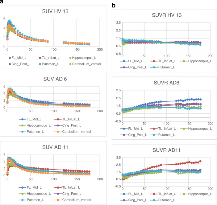
Methods: [18F] JNJ-64326067 PET scans were acquired for 90 min and then from 120 to 180 min in 5 participants with [18F]-florbetapir PET amyloid positive probable AD (73 ± 9 years) and 5 [18F]-florbetapir PET amyloid negative healthy controls (71 ± 7 years). Whole-body [18F] JNJ-64326067 PET CT scans were acquired in six healthy subjects for 5.5 h in 3 scanning sessions. Brain PET scans were visually reviewed. Regional quantification included kinetic analysis of distribution volume ration (DVR) estimated by Logan graphical analysis over the entire scan and static analysis of SUVr in late frames. Both methods used ventral cerebellar cortex as a reference region.
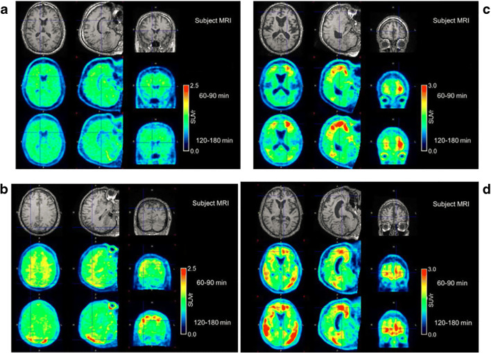
Results: One of the healthy controls had focal areas of PET signal in occipital and parietal cortex underlying the site of a gunshot injury as an adolescent; the other four healthy subjects had no tau brain signal. Four of the 5 AD participants had visually apparent retention of [18F] JNJ-64326067 in relevant cortical regions. One of the AD subjects was visually negative. Cortical signal in visually positive subjects approached steady state by 120 min. Temporal and frontal cortical SUVr/DVR values in visually positive AD subjects ranged from 1.21 to 3.09/1.2 to 2.18 and from 0.92 to 1.28/0.91 to 1.16 in healthy controls. Whole-body effective dose was estimated to be 0.0257 mSv/MBq for females and 0.0254 mSv/MBq for males.
Conclusions: [18F] JNJ-64326067 could be useful for detection and quantitation of tau aggregates.
Keywords: Alzheimer’s disease; Isoquinoline; PET; Tau aggregates.
Conflict of interest statement
Mark E. Schmidt, Luc Janssens, Diederik Moechars, Frederik J.R. Rombouts, Maarten Timmers, and Hartmuth Kolb are all full time employees of Janssen Research & Development, which provided funding and oversight of these studies and supported the discovery and preclinical characterization of JNJ-6436067. Olivier Barret, Cristian C. Constantinescu, Jennifer Madonia, David S. Russell, and Christine M. Sandiego are or were (OB, JM) full time employees of Invicro, a Konica Minolta company, New Haven, CT, USA, at the time of these studies.
Figures


References
-
- Chien DT, Bahri S, Szardenings AK, Walsh JC, Mu F, Su MY, et al. Early clinical PET imaging results with the novel PHF-tau radioligand [F-18]-T807. J Alzheimers Dis. 2013;34(2):457–468. - PubMed
-
- Ariza M, Kolb HC, Moechars D, Rombouts F, Andres JI. Tau positron emission tomography (PET) imaging: past, present, and future. J Med Chem. 2015;58(11):4365–4382. - PubMed
MeSH terms
Substances
LinkOut - more resources
Full Text Sources
Medical
