Review
. 2020 Dec;28(12):1022-1033.
doi: 10.1016/j.tim.2020.05.009. Epub 2020 Jun 11.

Double-Membrane Vesicles as Platforms for Viral Replication

Affiliations
Review

Double-Membrane Vesicles as Platforms for Viral Replication

Georg Wolff et al. Trends Microbiol. 2020 Dec.

Abstract

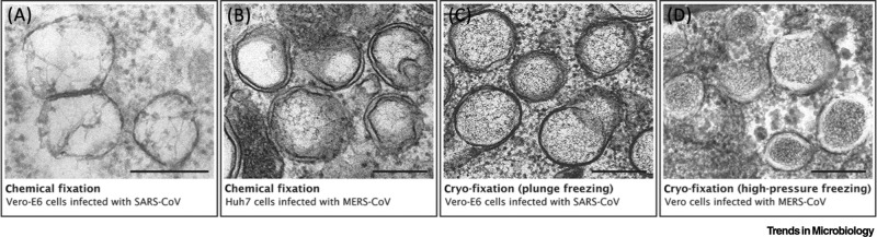
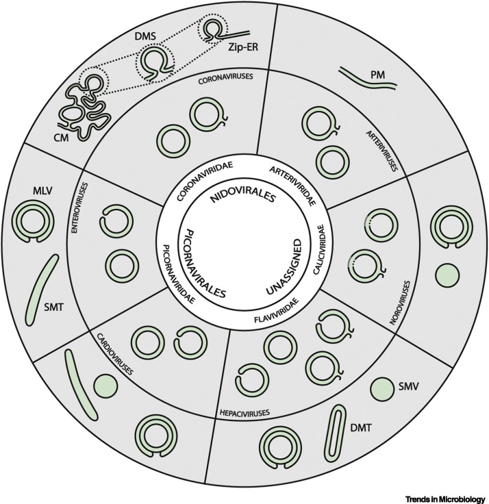
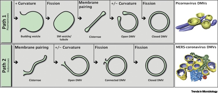
Viruses, as obligate intracellular parasites, exploit cellular pathways and resources in a variety of fascinating ways. A striking example of this is the remodelling of intracellular membranes into specialized structures that support the replication of positive-sense ssRNA (+RNA) viruses infecting eukaryotes. These distinct forms of virus-induced structures include double-membrane vesicles (DMVs), found during viral infections as diverse and notorious as those of coronaviruses, enteroviruses, noroviruses, or hepatitis C virus. Our understanding of these DMVs has evolved over the past 15 years thanks to advances in imaging techniques and modern molecular biology tools. In this article, we review contemporary understanding of the biogenesis, structure, and function of virus-induced DMVs as well as the open questions posed by these intriguing structures.

Keywords: DMV; endomembranes; membrane remodelling; positive-sense RNA viruses; replication membrane; viral replication organelles.


Figures

Figure I
Figure I
The Appearance of Double-Membrane Vesicles (DMVs) Can Drastically Change with Different Preparation Protocols for Electron Microscopy (EM). The changing appearance of DMVs in different preparation protocols illustrated by images of coronavirus-induced DMVs. (A,B) Examples of DMVs found in cells infected with severe acute respiratory syndrome (SARS)-coronavirus (A) and Middle East respiratory syndrome (MERS)-coronavirus (B), both prepared using chemical fixation but with different protocols. The protocol used in (A) results in poor membrane preservation, and the DMV morphology is difficult to interpret. In (B), both DMV membranes are readily visible, though large artefactual gaps between the inner and outer membrane are present. In general, chemically fixed DMVs are deformed and only roughly spherical. (C,D) Examples of cryo-fixed DMVs induced by SARS-coronavirus (C, fixed by plunge freezing), and MERS-coronavirus (D, fixed by high-pressure freezing), respectively. DMV membranes appear tightly apposed and with minimal deformation, illustrating the superior structural preservation of this approach. (D) High-pressure freezing is considered to be the optimal fixation method for room temperature EM [95] and reveals a core of dense material inside the DMVs, which are surrounded by a denser cytosol. (A) was adapted from [96], copyright © American Society for Microbiology, and (C) was adapted from [15]. (B) and (D) are unpublished images from samples described in [17,16], respectively. Scale bars, 250 nm.
Figure 1
Figure 1
Schematic Representing the Membrane Modifications Induced by Double-Membrane Vesicle (DMV)-Inducing Viruses. The viral order and family are indicated in the core and inner ring respectively. The next ring depicts the types of DMV architecture that have been described for different virus families, which include open and/or closed DMVs that may be disconnected or continuous with other structures as shown. In most cases, these connections are established with the endoplasmic reticulum (ER), but nidoviral DMVs are also interconnected and connected to other virus-induced membrane structures. For noroviruses the open/closed state of DMVs is unreported, indicated by broken lines. The outer ring depicts additional virus-induced membrane structures. Single-membrane vesicles (SMVs) have been documented for HCV and noroviruses [11,12,25,27]. Single-membrane tubules (SMTs), sometimes together with SMVs, appear early in picornavirus infections and are DMV precursors [8., 9., 10.,98]. Picornaviruses, HCV, and noroviruses also generate multilamellar vesicles (MLVs), which arise late in infection from massive enwrapping of certain replication organelle (RO) elements by others [8., 9., 10., 11., 12.]. Double-membrane tubules (DMTs) are also late structures that may arise from DMVs [12]. Nidoviruses induce additional double-membrane structures, including paired membranes (PMs) [13,29] which, in the case of coronaviruses, adopt different configurations, from a nonbranching form, termed zippered ER (Zip-ER) [20,99], to a highly labyrinthine structure known as convoluted membranes (CMs) [15., 16., 17.,100]. Small open double-membrane spherules (DMSs), highly reminiscent of the invaginated spherules induced by other +RNA viruses, are formed as invaginations in Zip-ER or CM [17,20,99]. Colours indicate cytosolic (grey) or luminal (green) space.
Figure 2
Figure 2
Pathways of Double-Membrane Vesicle (DMV) Biogenesis and Representative 3D Models. The biogenesis of DMVs induced by different +RNA viruses appear to occur through two main pathways. (Path 1, top) Initially, by the induction of positive membrane curvature, single-membrane (SM) vesicles or tubules bud out from the donor organelle. Through membrane pairing, these structures form cisternae that curve (inducing positive and negative curvature at the outer and inner membrane, respectively) to finally seal and transform into a closed DMV. This mechanism is supported by intermediate structures found in picornavirus infections [8., 9., 10.] and it has been speculated that norovirus- and Hepatitis C (HCV)-induced DMVs may be formed in a similar way [11,27,28]. (Top right) A 3D model of DMVs (yellow) next to virus-induced SM tubules (blue) in cells infected with encephalomyocarditis virus (family Picornaviridae, genus Cardiovirus), adapted from [10]. (Path 2, bottom) A different pathway of DMV biogenesis appears to take place in nidovirus-infected cells [29., 30., 31.]. Here, endoplasmic reticulum (ER) membranes would pair to form cisternae; these would subsequently curve and undergo one or two fission events to result in connected or free-floating DMVs. (Bottom right) A 3D model of Middle East respiratory syndrome (MERS)-coronavirus-induced DMVs (yellow and lilac, outer and inner membrane, respectively), connected to the ER (green) and convoluted membranes (blue) that include a double-membrane spherule (orange). Model adapted from [17].

References

    1. Scutigliani E.M., Kikkert M. Interaction of the innate immune system with positive-strand RNA virus replication organelles. Cytokine Growth Factor Rev. 2017;37:17–27. - PMC - PubMed
    1. de Castro I.F. Virus factories: biogenesis and structural design. Cell. Microbiol. 2013;15:24–34. - PMC - PubMed
    1. Liu L. From crescent to mature virion: vaccinia virus assembly and maturation. Viruses. 2014;6:3787–3808. - PMC - PubMed
    1. Tenorio R. Function, architecture, and biogenesis of reovirus replication neoorganelles. Viruses. 2019;11:288. - PMC - PubMed
    1. Harak C., Lohmann V. Ultrastructure of the replication sites of positive-strand RNA viruses. Virology. 2015;479:418–433. - PMC - PubMed