

Hemodynamic and Histopathological Changes in the Early Phase of the Development of an Intracranial Aneurysm
- PMID: 32536660
- PMCID: PMC7358784
- DOI: 10.2176/nmc.st.2020-0072
Hemodynamic and Histopathological Changes in the Early Phase of the Development of an Intracranial Aneurysm
Abstract
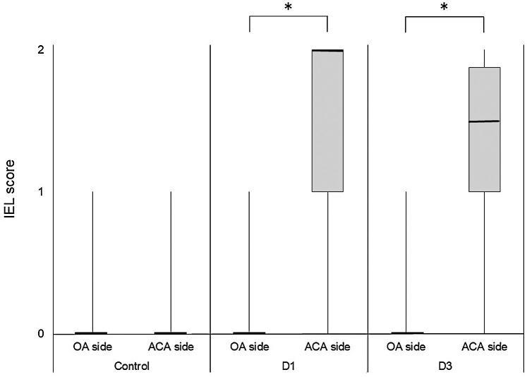
Hemodynamic stress and chronic inflammation are closely associated with the pathogenesis of intracranial aneurysms (IAs). However, the hemodynamic and biological mechanisms triggering IA formation remain to be elucidated. To clarify them, computational fluid dynamics (CFD) and histopathological analyses in the early phase of IA development using an experimentally induced IA model in rats were conducted. Histological changes in the early phase of IA development were observed under a scanning electron microscope (SEM) and a transmission electron microscope (TEM). Using data from 7-T magnetic resonance angiography (7T-MRA), CFD analyses were performed to determine wall shear stress (WSS) and wall pressure (WP) at the prospective site of IA. A bump-like protrusion named an "intimal pad" was located in the anterior cerebral artery (ACA) immediately distal to the apex of the bifurcation. TEM showed the degeneration of the internal elastic lamina (IEL) and longitudinally elongated smooth muscle cells (SMCs) that switched from the contractile to the proliferative phenotype and penetrated between two divided layers of the degenerated IEL in the prospective site of the IA. However, no inflammatory cells were observed. CFD analyses showed no particular pattern of WSS and WP at the prospective IA site. IEL degeneration and the phenotypic change and longitudinal elongation of SMCs were identified as the early events in IA development. CFD analyses and TEM data suggest that these biological events may be derived from increased circumferential wall stress due to increased blood pressure and increased longitudinal wall strain due to the existence of the intimal pad.
Keywords: hemodynamics; internal elastic lamina; intracranial aneurysm; smooth muscle cell.
Conflict of interest statement
The authors have no personal financial or institutional interest in any of the drugs, materials, or devices in the article. H.K., T.I., K.Y., and S.M are members of The Japan Neurosurgical Society (JNS) and have registered online Self-reported COI Disclosure Statement Forms through the website for JNS members.
Figures


References
MeSH terms
LinkOut - more resources
Full Text Sources
Medical
Research Materials
Miscellaneous