

Human adipose-derived stromal vascular fraction: characterization, safety and therapeutic potential in an experimental mouse model of articular injury
- PMID: 32536767
- PMCID: PMC7282273
- DOI: 10.46582/jsrm.1601004
Human adipose-derived stromal vascular fraction: characterization, safety and therapeutic potential in an experimental mouse model of articular injury
Abstract
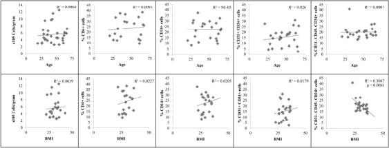
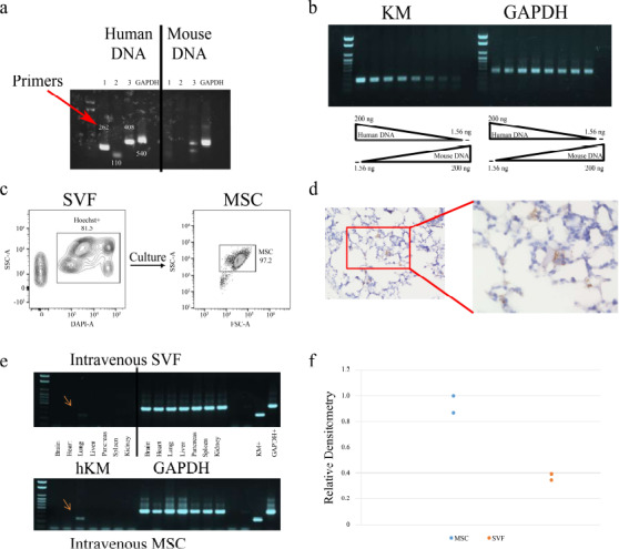
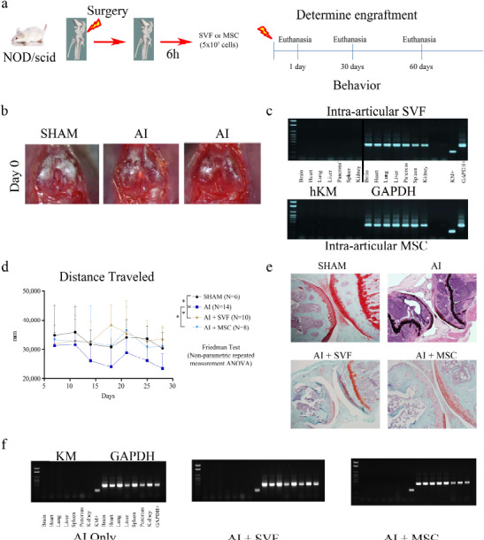
Due to their capacity to self-renew, proliferate and generate multi-lineage cells, adult-derived stem cells offer great potential in regenerative therapies to treat maladies such as diabetes, cardiac disease, neurological disorders and orthopedic injuries. Commonly derived from adipose tissue, the stromal vascular fraction (SVF), a heterogeneous cell population enriched with mesenchymal stem cells (MSCs), has garnered interest as a cellular therapy due to ease of accessibility as an autologous, point-of-care application. However, the heterogeneous cell population within SVF is not historically taken into consideration when injecting into patients. Here, we characterized SVF, determined its safety and verify its therapeutic effects in a NOD/scid mouse model of articular injury. SVF were isolated from lipoaspirates utilizing a commercially available system (InGeneron Inc.), while MSCs were isolated from SVF via cell culture. Flow cytometry showed that neither age nor BMI affects the frequency of progenitor cells-like (CD31+CD34+), immune cells-like (CD4+) T cells, (CD14+) monocytes and total number of cells obtained. However, there was a negative correlation between donor BMI and MSC frequency within the SVF. ELISAs showed that following LPS activation in SVF, there were low levels of TNF-α and high levels of IL-10 secreted. However, T cell activation with anti-CD3 or anti-CD3+ anti-CD28, while leading to expected high levels of IFN-γ, did not lead to significant levels of TGF-β. PCR analysis showed no significant numbers of cells outside the joint 1-hour post injection, moreover, no engraftment or abnormal growth in other organs 60-days post injection. Finally, both cell populations were able to ameliorate disease progression, as confirmed by the increase in movement of treated groups compared to injured groups. Noteworthy, the histological analysis indicated that there was no cartilage growth, suggesting an alternative therapeutic mechanism to cartilage regeneration.
Keywords: Adipose; Autologous stem cells; Cartilage; Mesenchymal stem cell.
Copyright © Journal of Stem Cells and Regenerative Medicine.
Conflict of interest statement
None
Figures

Similar articles
-
The ratio of ADSCs to HSC-progenitors in adipose tissue derived SVF may provide the key to predict the outcome of stem-cell therapy.Clin Transl Med. 2018 Feb 7;7(1):5. doi: 10.1186/s40169-018-0183-8. Clin Transl Med. 2018. PMID: 29417261 Free PMC article.
-
Characterization of adipose tissue-derived stromal vascular fraction for clinical application to cartilage regeneration.In Vitro Cell Dev Biol Anim. 2015 Feb;51(2):142-50. doi: 10.1007/s11626-014-9814-6. Epub 2014 Nov 1. In Vitro Cell Dev Biol Anim. 2015. PMID: 25361717
-
Concise Review: Fat and Furious: Harnessing the Full Potential of Adipose-Derived Stromal Vascular Fraction.Stem Cells Transl Med. 2017 Apr;6(4):1096-1108. doi: 10.1002/sctm.16-0337. Epub 2017 Jan 6. Stem Cells Transl Med. 2017. PMID: 28186685 Free PMC article. Review.
-
Comparative Efficacy of Autologous Stromal Vascular Fraction and Autologous Adipose-Derived Mesenchymal Stem Cells Combined With Hyaluronic Acid for the Treatment of Sheep Osteoarthritis.Cell Transplant. 2018 Jul;27(7):1111-1125. doi: 10.1177/0963689718773333. Epub 2018 Jun 18. Cell Transplant. 2018. PMID: 29909687 Free PMC article.
-
Autologous and Allogenic Utilization of Stromal Vascular Fraction and Decellularized Extracellular Matrices in Orthopedic Surgery: A Scoping Review.Arch Bone Jt Surg. 2022 Oct;10(10):827-832. doi: 10.22038/ABJS.2022.59635.2943. Arch Bone Jt Surg. 2022. PMID: 36452418 Free PMC article.
Cited by
-
Vasculogenic potential of adipose tissue derived stem cells from patients with chronic spinal cord injury and pressure injuries.Angiogenesis. 2025 Sep 10;28(4):48. doi: 10.1007/s10456-025-10002-y. Angiogenesis. 2025. PMID: 40928671 Free PMC article.
-
Why and how to use the body's own stem cells for regeneration in musculoskeletal disorders: a primer.J Orthop Surg Res. 2022 Jan 21;17(1):36. doi: 10.1186/s13018-022-02918-8. J Orthop Surg Res. 2022. PMID: 35062984 Free PMC article.
-
Heterogeneous Cells as well as Adipose-Derived Stromal Cells in Stromal Vascular Fraction Contribute to Enhance Anabolic and Inhibit Catabolic Factors in Osteoarthritis.Stem Cell Rev Rep. 2023 Oct;19(7):2407-2419. doi: 10.1007/s12015-023-10589-z. Epub 2023 Jul 21. Stem Cell Rev Rep. 2023. PMID: 37477775
-
Regeneration of Biomechanically Functional Tendon Tissue Following Injection of Uncultured, Autologous, Adipose-Derived Regenerative Cells into Partial Achilles Tendon Defects in Rabbits.Int J Mol Sci. 2025 Jul 16;26(14):6800. doi: 10.3390/ijms26146800. Int J Mol Sci. 2025. PMID: 40725049 Free PMC article.
-
Custom tailoring of Cell therapies to address cartilage damages efficiently.J Stem Cells Regen Med. 2020 May 27;16(1):1-2. doi: 10.46582/jsrm.1601001. eCollection 2020. J Stem Cells Regen Med. 2020. PMID: 32536764 Free PMC article. No abstract available.
References
-
- Rehman J, Traktuev D, Li J, Merfeld-Clauss S, Temm-Grove CJ, Bovenkerk JE, Pell CL, Johnstone BH, Considine RV, March KL. Secretion of angiogenic and antiapoptotic factors by human adipose stromal cells. Circulation. 2004;109((10)):1292–8. - PubMed
-
- Traktuev DO, Prater DN, Merfeld-Clauss S, Sanjeevaiah AR, Saadatzadeh MR, Murphy M, Johnstone BH, Ingram DA, March KL. Robust functional vascular network formation in vivo by cooperation of adipose progenitor and endothelial cells. Circ Res. 2009;104((12)):1410–20. - PubMed
-
- Zuk PA, Zhu M, Mizuno H, Huang J, Futrell JW, Katz AJ, Benhaim P, Lorenz HP, Hedrick MH. Multilineage cells from human adipose tissue: implications for cell-based therapies. Tissue Eng. 2001;7((2)):211–28. - PubMed
-
- Strioga M, Viswanathan S, Darinskas A, Slaby O, Michalek J. Same or not the same? Comparison of adipose tissue-derived versus bone marrow-derived mesenchymal stem and stromal cells. Stem Cells Dev. 2012;21((14)):2724–52. - PubMed
Grants and funding
LinkOut - more resources
Full Text Sources
Research Materials