

Achatina fulica mucous improves cell viability and increases collagen deposition in UVB-irradiated human fibroblast culture
- PMID: 32536768
- PMCID: PMC7282269
- DOI: 10.46582/jsrm.1601005
Achatina fulica mucous improves cell viability and increases collagen deposition in UVB-irradiated human fibroblast culture
Abstract
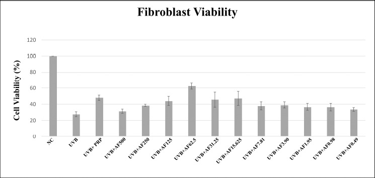
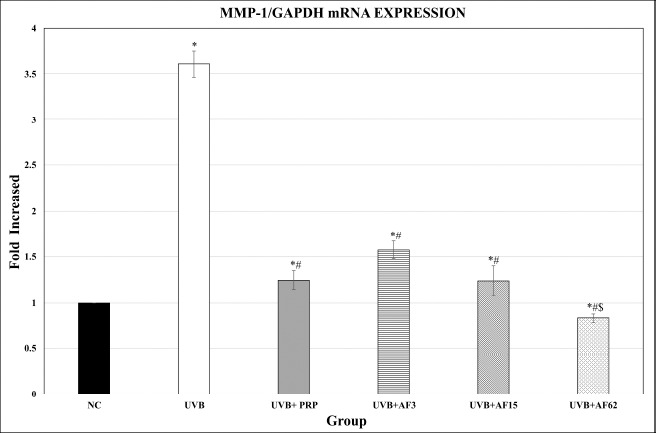
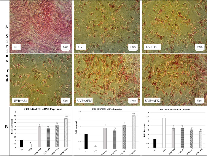
Introduction: Ultraviolet radiation induces skin photoaging by increasing matrix metalloproteinase-1 (MMP-1). MMP-1 degrades type I and III collagen that comprise the dermal connective tissue. Achatina fulica mucous (AFM) is a natural remedy that has protective effects on fibroblasts and collagen. Objective: To investigate the effects of AFM on cell viability and collagen deposition in UVB-irradiated human fibroblast culture. Methods: The mucous was extracted from 50 Achatina fulica snails that were stimulated by a 5-10 Volt electricity shock for 30-60 seconds and converted into powder by the freeze-drying process. The human dermal fibroblast culture was divided into six groups: group 1 were normal fibroblasts without UVB irradiation as normal control, groups 2-5 consisted of 100 mJ/cm2 UVB-irradiated fibroblasts. Group 2 had no treatment as negative control, group 3 was treated by PRP 10% as positive control group and groups 4-6 were treated by various concentrations of AFM (3.9; 15.625 and 62.5 μg/mL). At the end of the experiment, the proliferation was assessed with MTT assay, furthermore collagen deposition was measured by Sirius red assay. Real Time-PCR (RT-PCR) was performed to quantify Coll I, Coll III and MMP-1 mRNA expression, then to measured COL 1/COL III ratio. Results: UVB induced significant lower viability, upregulated MMP-1 and downregulated COL I and COL III mRNA expressions. Meanwhile AFM treated groups demonstrated higher cell viability with downregulation of MMP-1 and upregulation of COL I and COL III mRNA expressions. The ratio of COL I/ III expression was significantly (p<0.05) lower in the AFM treated groups compared to the UVB group. Among AFM treated groups, administration of 62.5 μg/mL AFM represented the best result. Conclusion: AFM may ameliorate viability of UVB-irradiated human fibroblast culture which associates with downregulating MMP-1, upregulating COL I and Col III, and reducing COL I/III ratio.
Keywords: Achatina fulica; Collagen; MMP-1; UVB-irradiated human fibroblast culture; Viability.
Copyright © Journal of Stem Cells and Regenerative Medicine.
Conflict of interest statement
This work was supported by a grant from Ministry of Research, Technology and Higher Education of the Republic of Indonesia through Beasiswa Unggulan Dosen Indonesia Dalam Negeri (BUDI-DN) for the Ph.D. scholarship program (number PRJ-1375/LPDP.4 /2016).
Figures
Similar articles
-
UVB-irradiated human keratinocytes and interleukin-1alpha indirectly increase MAP kinase/AP-1 activation and MMP-1 production in UVA-irradiated dermal fibroblasts.Chin Med J (Engl). 2006 May 20;119(10):827-31. Chin Med J (Engl). 2006. PMID: 16732985
-
IL-1 receptor antagonist attenuates MAP kinase/AP-1 activation and MMP1 expression in UVA-irradiated human fibroblasts induced by culture medium from UVB-irradiated human skin keratinocytes.Int J Mol Med. 2005 Dec;16(6):1117-24. Int J Mol Med. 2005. PMID: 16273295
-
Anti-photoaging potential of Botulinum Toxin Type A in UVB-induced premature senescence of human dermal fibroblasts in vitro through decreasing senescence-related proteins.J Photochem Photobiol B. 2014 Apr 5;133:115-23. doi: 10.1016/j.jphotobiol.2014.03.009. Epub 2014 Mar 25. J Photochem Photobiol B. 2014. PMID: 24727404
-
The effect of narrowband ultraviolet B on the expression of matrix metalloproteinase-1, transforming growth factor-beta1 and type I collagen in human skin fibroblasts.Clin Exp Dermatol. 2007 Mar;32(2):180-5. doi: 10.1111/j.1365-2230.2006.02309.x. Epub 2006 Nov 27. Clin Exp Dermatol. 2007. PMID: 17137474
-
Increase of collagen synthesis by obovatol through stimulation of the TGF-beta signaling and inhibition of matrix metalloproteinase in UVB-irradiated human fibroblast.J Dermatol Sci. 2007 May;46(2):127-37. doi: 10.1016/j.jdermsci.2007.02.001. Epub 2007 Mar 7. J Dermatol Sci. 2007. PMID: 17346934
Cited by
-
Understanding Snail Mucus Biosynthesis and Shell Biomineralisation through Genomic Data Mining of the Reconstructed Carbohydrate and Glycan Metabolic Pathways of the Giant African Snail (Achatina fulica).Biology (Basel). 2023 Jun 9;12(6):836. doi: 10.3390/biology12060836. Biology (Basel). 2023. PMID: 37372121 Free PMC article.
-
Synergistic Antibacterial Effect of Mucus Fraction from Cornu aspersum and Cirpofloxacin Against Pathogenic Bacteria Isolated from Wounds of Diabetic Patients.Antibiotics (Basel). 2025 Mar 4;14(3):260. doi: 10.3390/antibiotics14030260. Antibiotics (Basel). 2025. PMID: 40149071 Free PMC article.
-
Recent advances in exosomal non-coding RNA-based therapeutic approaches for photoaging.Skin Res Technol. 2023 Sep;29(9):e13463. doi: 10.1111/srt.13463. Skin Res Technol. 2023. PMID: 37753673 Free PMC article. Review.
-
Puerarin Reduces Oxidative Damage and Photoaging Caused by UVA Radiation in Human Fibroblasts by Regulating Nrf2 and MAPK Signaling Pathways.Nutrients. 2022 Nov 9;14(22):4724. doi: 10.3390/nu14224724. Nutrients. 2022. PMID: 36432411 Free PMC article.
-
Anti-Photoaging Effect of Rhodiola rosea Fermented by Lactobacillus plantarum on UVA-Damaged Fibroblasts.Nutrients. 2022 Jun 1;14(11):2324. doi: 10.3390/nu14112324. Nutrients. 2022. PMID: 35684124 Free PMC article.
References
-
- Cavinato M, Jansen-Dürr P. Molecular mechanisms of UVB-induced senescence of dermal fibroblasts and its relevance for photoaging of the human skin. Exp Gerontol. 2017;94:78–82. - PubMed
-
- Scharffetter—Kochanek K, Brenneisen P, Wenk J, Herrmann G, Ma W, Kuhr L et al. Photoaging of the skin from phenotype to mechanisms. Exp Gerontol. 2000;35((3)):307–316. - PubMed
-
- Tigges J, Krutmann J, Fritsche E, Haendeler J, Schaal H, Fischer JW et al. The hallmarks of fibroblast ageing. Mech Ageing Dev. 2014;138:26–44. - PubMed
-
- Peng Y, Song X, Zheng Y, Wang X, Lai W. Circular RNA profiling reveals that circCOL3A1-859267 regulate type I collagen expression in photoaged human dermal fibroblasts. Biochem Biophys Res Commun. 2017;486((2)):277–84. - PubMed
LinkOut - more resources
Full Text Sources
Research Materials
Miscellaneous