Review
. 2020 May 27:11:853.
doi: 10.3389/fimmu.2020.00853. eCollection 2020.

Diverse Virus and Host-Dependent Mechanisms Influence the Systemic and Intrahepatic Immune Responses in the Woodchuck Model of Hepatitis B

Affiliations
Review

Diverse Virus and Host-Dependent Mechanisms Influence the Systemic and Intrahepatic Immune Responses in the Woodchuck Model of Hepatitis B

Tomasz I Michalak. Front Immunol. .

Abstract

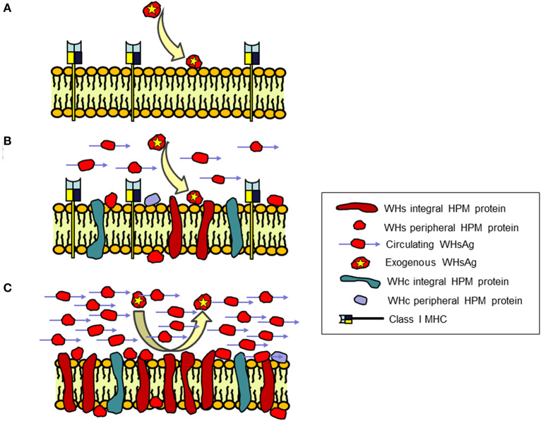
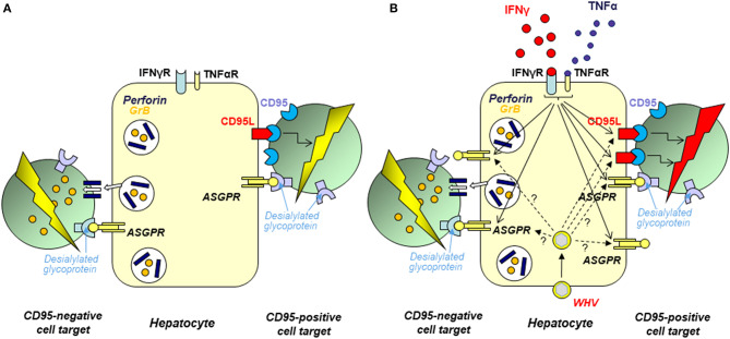
Woodchuck infected with woodchuck hepatitis virus (WHV) represents the pathogenically nearest model of hepatitis B and associated hepatocellular carcinoma (HCC). This naturally occurring animal model also is highly valuable for development and preclinical evaluation of new anti-HBV agents and immunotherapies against chronic hepatitis (CH) B and HCC. Studies in this system uncovered a number of molecular and immunological processes which contribute or likely contribute to the immunopathogenesis of liver disease and modulation of the systemic and intrahepatic innate and adaptive immune responses during hepadnaviral infection. Among them, inhibition of presentation of the class I major histocompatibility complex on chronically infected hepatocytes and a role of WHV envelope proteins in this process, as well as augmented hepatocyte cytotoxicity mediated by constitutively expressed components of CD95 (Fas) ligand- and perforin-dependent pathways, capable of eliminating cells brought to contact with hepatocyte surface, including activated T lymphocytes, were uncovered. Other findings pointed to a role of autoimmune response against hepatocyte asialoglycoprotein receptor in augmenting severity of liver damage in hepadnaviral CH. It was also documented that WHV in the first few hours activates intrahepatic innate immunity that transiently decreases hepatic virus load. However, this activation is not translated in a timely manner to induction of virus-specific T cell response which appears to be hindered by defective activation of antigen presenting cells and presentation of viral epitopes to T cells. The early WHV infection also induces generalized polyclonal activation of T cells that precedes emergence of virus-specific T lymphocyte reactivity. The combination of these mechanisms hinder recognition of virus allowing its dissemination in the initial, asymptomatic stages of infection before adaptive cellular response became apparent. This review will highlight a range of diverse mechanisms uncovered in the woodchuck model which affect effectiveness of the anti-viral systemic and intrahepatic immune responses, and modify liver disease outcomes. Further exploration of these and other mechanisms, either already discovered or yet unknown, and their interactions should bring more comprehensive understanding of HBV pathogenesis and help to identify novel targets for therapeutic and preventive interventions. The woodchuck model is uniquely positioned to further contribute to these advances.

Keywords: asialoglycoprotein receptor; hepatocyte as cytotoxic immune effector; intrahepatic innate and adaptive immune responses; major histocompatibility complex presentation; pre-acute infection; toll-like receptors; virus-hepatocyte interaction; woodchuck model of hepatitis B.


Figures

Figure 1
Figure 1
Schematic presentation of serum immunovirological and molecular profiles of occult infections caused by WHV. (A) Secondary occult infection (SOI) continuing after resolution of self-limited acute hepatitis caused by a liver pathogenic dose greater or equal to 1 × 10³ WHV characterized by persistent asymptomatic infection negative for serum WHsAg but reactive for anti-WHc antibodies, positive or negative for anti-WHs antibodies, and positive for WHV DNA at levels below 100–200 virus copies/mL. (B) Primary occult infection (POI) developing after exposure to a liver non-pathogenic dose of <1 × 10³ WHV virions that persists as a seronegative infection but remains serum WHV DNA reactive at levels usually below 100–200 virus copies/mL.
Figure 2
Figure 2
Abbreviated concept of the development of occult and overt (symptomatic) WHV infections, sites of replicating virus, and serological appearance and infection outcomes in relation to a dose of invading virus. For details see text.
Figure 3
Figure 3
Outline of the study investigating virological, immunological and liver injury outcomes in woodchucks injected with multiple liver-nonpathogenic doses of WHV. (A) Four WHV-naïve, healthy animals were intravenously injected with 6 weekly doses of 110 WHV virions (Phase 1). Six months later they were challenged with another 6 weekly doses of 110 virions each of the same virus (Phase 2). The total quantity of the virus administered was 1,320 virions and cumulatively exceeded a liver pathogenic dose of 1 × 10³ virions. Three-and-half months later, the animals were re-challenged with either a liver pathogenic dose of 1.1 × 10⁶ virions of the same virus or injected with sterile PBS as controls (Challenge). (B) An example of the outcomes investigated in a woodchuck subjected to two rounds of injections with 6 weekly 110-viron doses of WHV and then injected with PBS. The data showed the lack of appearance of serologically detectable infection, including absence of anti-WHc antibodies which are very sensitive indicator of exposure to WHV, or hepatitis (upper panel). WHV-specific T cell response (middle panel) was preceded by ConA-induced, generalized T cell proliferation (lower panel). For details and abbreviations, see the legend to Figure 4. Figures adopted with modifications from Gujar et al. (52).
Figure 4
Figure 4
Distinct kinetics of WHV-specific and generalized T cell proliferative response following infection with a liver pathogenic dose of WHV and subsequent challenge with the same ioclum. (A) Schematic depiction of the cumulative data on discordance between virus-specific and generalized, mitogen-induced T cell proliferative responses. The profiles were compiled based on the data obtained from 4 animals with self-limited acute hepatitis after infection with 1.9 × 10¹¹ DNase digestion-protected WHV virions using a flow cytometry CFSE assay to assess T cell proliferation in response to five different recombinant or native WHV proteins and WHc97−110 peptide (stimulation index values, SI) and in response to five 2-fold dilutions of mitogen concanavalin A (ConA) ranging from 1.25 to 20 μg/mL measured by a [3H]-adenine incorporation assay (mean mitogen stimulation index values, MMSI). The mean of the highest SI given by any WHV antigen or that of MMSI in response to any ConA concentration from each of four animals were used to construct the profiles. Solid red upward arrowheads indicate injections with WHV. Black and red stars mark peaks of mitogen-induced and WHV-specific T cell responses, respectively. The data demonstrate that WHV first triggers polyclonal generalized activation of T cells and then WHV-specific T cell response. (B) An example of the WHV-specific T cell response to recombinant WHV e protein (rWHe) in the same 4 animals shown in panel A. (C) An example of the mitogen-induced T cell proliferation after infection and challenge with WHV in response to stimulation with ConA. Figures adopted with modifications from Gujar et al. (53).
Figure 5
Figure 5
Schematic presentation of the concept of an immune protective barrier at the surface of hepatocytes in chronic WHV infection created by virus envelope (WHs) proteins associated with hepatocyte plasma membranes (HPM) that coincides with depletion of class I MHC on the membranes. HPM from (A) Healthy, WHV-negative woodchucks; (B) WHV-infected animals with acute hepatitis (AH), and (C) WHV-infected animals with chronic hepatitis (CH). CH in contrast to AH is characterized by irreversible incorporation of large (saturating) quantities of WHs proteins into HPM lipid bilayer and presence of WHs reactivity loosely associated with HPM structure behaving as peripheral membrane protein. In addition, CH occurs in the context of large quantities of circulating virus envelope proteins carrying WHs antigenic specificity (WHsAg) and inability of HPM to bind exogenous purified WHsAg in contrast to HPM from healthy woodchucks and animals with AH. WHV core (WHc) proteins are also incorporated as integral and joined with HPM as peripheral membrane proteins, however there are no differences between AH and CH in their HPM quantities and susceptibility to removal by agents dissociating HPM. In addition, HPM from CH are voided of class I MHC presence in contrast to HPM from acutely infected and healthy animals. Together, the concept assumes that WHs proteins incorporate at saturating quantities into hepatocyte surface membranes in combination with large amounts of circulating WHsAg and absence of class I MHC presentation at the membranes form an immune protective barrier that shelters infected hepatocytes from immune elimination. For details see Michalak and Churchill (115), Michalak et al. (116, 117), Michalak and Lin (118), Michalak et al. (119), and Wang and Michalak (120).
Figure 6
Figure 6
Schematic presentation of the mechanisms and factors affecting elimination of cells brought to contact with hepatocyte surface. In opposite to the previous opinion, hepatocytes intrinsically transcribe perforin, as well as CD95 and CD95 ligand (CD95L) and can utilize both perforin-granzyme B (GrB) and CD95L-CD95 pathways to kill other cells. The acquired experimental data indicate that hepatocytes can recognize other cells via interaction between desialylated glycoproteins on the target cell surface and asialoglycoprotein receptor (ASGPR) on hepatocyte plasma membrane. (A) Normal hepatocytes show ability to eliminate contacted cells by both CD95L- and perforin-dependent mechanisms. ASGPR involvement in this process is supported by demonstration that desialylation of surface glycoproteins on target cells enhanced their susceptibility to hepatocyte-mediated killing and that inhibition of hepatocyte ASGPR by asilofetuin, a ASGPR-specific ligand, or by silencing of ASGPR gene by specific siRNA significantly limited hepatocyte-facilitated cell killing. (B) The hepatocyte capacity to eliminate cells brought to contact with their surface is augmented after hepatocyte exposure to IFN-γ or TNF-α. This appears to be a consequence of the combined effects of enhanced expression of the cytotoxic effector molecules and augmented display of ASGPR on hepatocyte surface (indicated by arrows with continuous stamps). In chronic WHV hepatitis and resolved acute WHV infection, hepatocyte CD95L- and perforin-dependent cytotoxicity is augmented when compared to hepatocytes from livers of healthy woodchucks. This appears to be a consequence of liver inflammation caused by WHV which progression and resolution are associated with augmented production of many bioactive factors including IFN-γ and TNF-α. It also is possible that the augmented hepatocyte cytotoxicity in some situations could be due to a direct effect of virus (WHx protein) via upregulated expression of the cytotoxic effector molecules or ASGPR in hepatocytes (marked by arrows with dashed stamps). For details see Guy et al. (–151), Guy et al. (152).

References

    1. Summers J, Smolec JM, Snyder R. A virus similar to human hepatitis B virus associated with hepatitis and hepatoma in woodchucks. Proc Natl Acad Sci USA. (1978) 75:4533–7. 10.1073/pnas.75.9.4533 - DOI - PMC - PubMed
    1. Werner BG, Smolec JM, Snyder RL, Summers J. Serological relationship of woodchuck hepatitis virus to human hepatitis B virus. J Virol. (1979) 32:314–22. 10.1128/JVI.32.1.314-322.1979 - DOI - PMC - PubMed
    1. Summers J, Mason WS. Replication of the genome of a hepatitis B–like virus by reverse transcription of an RNA intermediate. Cell. (1982) 29:403–15. 10.1016/0092-8674(82)90157-X - DOI - PubMed
    1. Galibert F, Chen TN, Mandart E. Nucleotide sequence of a cloned woodchuck hepatitis virus genome: comparison with the hepatitis B virus sequence. J Virol. (1982) 41:51–65. 10.1128/JVI.41.1.51-65.1982 - DOI - PMC - PubMed
    1. Michalak TI. Occult persistence and lymphotropism of hepadnaviral infection: insights from the woodchuck viral hepatitis model. Immunol Rev. (2000) 174:98–111. 10.1034/j.1600-0528.2002.017406.x - DOI - PubMed

Publication types