. 2020 May 27:11:854.
doi: 10.3389/fimmu.2020.00854. eCollection 2020.

Analysis of the Targets and Glycosylation of Monoclonal IgAs From MGUS and Myeloma Patients

Affiliations

Analysis of the Targets and Glycosylation of Monoclonal IgAs From MGUS and Myeloma Patients

Adrien Bosseboeuf et al. Front Immunol. .

Abstract

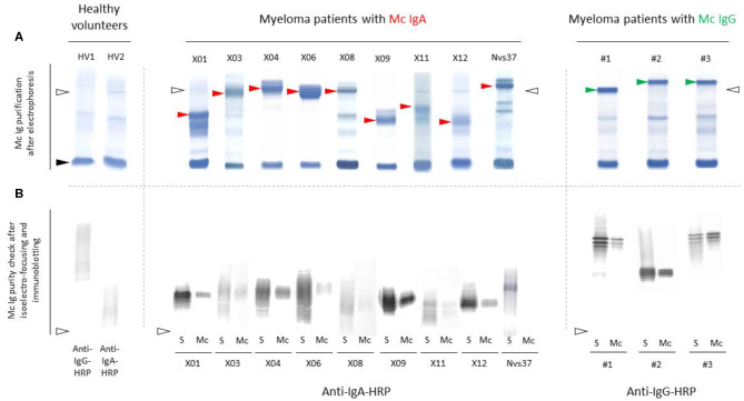
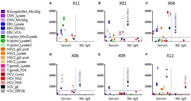
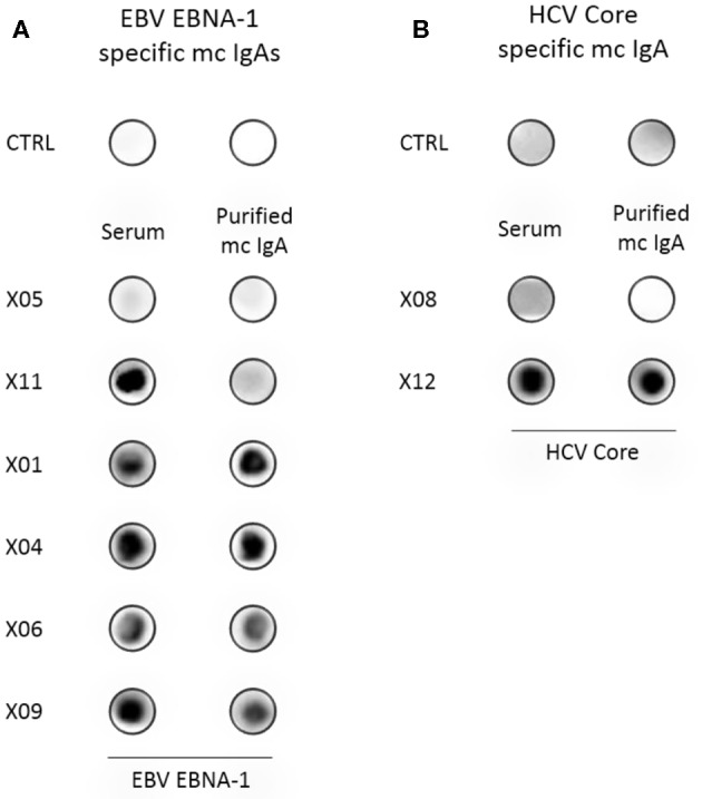
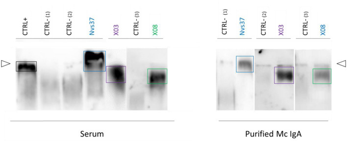
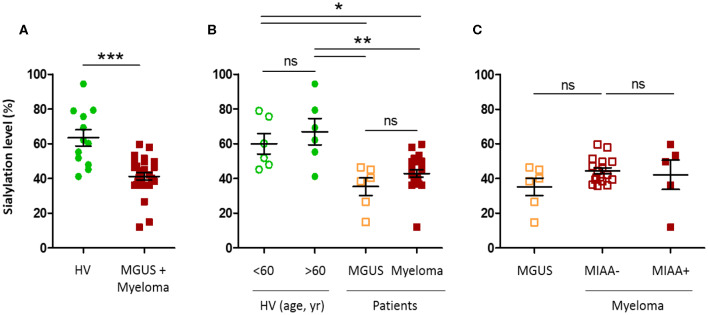
Previous studies showed that monoclonal immunoglobulins G (IgGs) of "monoclonal gammopathy of undetermined significance" (MGUS) and myeloma were hyposialylated, thus presumably pro-inflammatory, and for about half of patients, the target of the monoclonal IgG was either a virus-Epstein-Barr virus (EBV), other herpes viruses, hepatitis C virus (HCV)-or a glucolipid, lysoglucosylceramide (LGL1), suggesting antigen-driven disease in these patients. In the present study, we show that monoclonal IgAs share these characteristics. We collected 35 sera of patients with a monoclonal IgA (6 MGUS, 29 myeloma), and we were able to purify 25 of the 35 monoclonal IgAs (6 MGUS, 19 myeloma). Monoclonal IgAs from MGUS and myeloma patients were significantly less sialylated than IgAs from healthy volunteers. When purified monoclonal IgAs were tested against infectious pathogens and LGL1, five myeloma patients had a monoclonal IgA that specifically recognized viral proteins: the core protein of HCV in one case, EBV nuclear antigen 1 (EBNA-1) in four cases (21.1% of IgA myeloma). Monoclonal IgAs from three myeloma patients reacted against LGL1. In summary, monoclonal IgAs are hyposialylated and as described for IgG myeloma, significant subsets (8/19, or 42%) of patients with IgA myeloma may have viral or self (LGL1) antigen-driven disease.

Keywords: Epstein–Barr virus; hepatitis C virus; infectious antigens; lysoglucosylceramide (LGL-1); monoclonal gammopathy of undetermined significance (MGUS); monoclonal immunoglobulin A (IgA); multiple myeloma; sialylation.


Figures

Figure 1
Figure 1
Separation of monoclonal IgAs from other Igs in serum. (A) Monoclonal IgAs (red arrowheads) and monoclonal IgGs used as controls (green arrowheads) were separated from other Igs in serum as published (–11, 19). In the serum of healthy volunteers (HV), only polyclonal Igs are found; thus, no band is seen, only a smear. Note that monoclonal IgAs migrate in the β zone, are less well-separated than monoclonal IgGs, and thus are more difficult to purify than monoclonal IgGs. (B) The purity of monoclonal IgAs and IgGs was verified using isoelectric focusing (IEF) and immunoblotting. (A,B) Nine examples are shown for monoclonal IgAs and three for monoclonal IgGs (S, serum; Mc, monoclonal Ig separated from polyclonal Igs). Polyclonal Igs are represented by smears, whereas monoclonal Igs are represented by a single band in serum protein electrophoresis (A) and due to different migration according to different degrees of sialylation during IEF, as a stack of bands (B). The lines of sample deposit are indicated by white arrowheads. The albumin band is shown with a black arrowhead (A).
Figure 2
Figure 2
Viral targets of purified monoclonal IgAs from myeloma patients, as determined by the multiplexed infectious antigen micro-array (MIAA) revealed using a DylightTM 680-labeled goat anti-human IgA Fc antibody. For each patient, serum and purified monoclonal (Mc) IgA were incubated in parallel in the MIAA assay; results shown as fluorescent intensity represent either unseparated IgAs (left) or the patient's monoclonal IgA (right). (A) A patient with a Mc IgA that does not react with any pathogen of the MIAA. (B–E) Four patients with Epstein–Barr virus (EBV)-specific Mc IgAs. (F) One patient with a hepatitis C virus (HCV)-specific Mc IgA. EBV nuclear antigen (EBNA-1) signals are shown in dark blue dots, HCV core signals in red dots, and positive thresholds are shown in dotted lines. (A) For patient X11, the serum contained IgAs that recognized Borrelia burgdorferi, EBV EBNA-1, EBV VCA, Helicobacter pylori lysates 1 and 2, HCV NS3, and varicella zoster virus (VZV) ORF26 protein, whereas the purified Mc IgA did not recognize anything on the MIAA array. (B) For patient X01, the serum contained IgAs that recognized a mix of cytomegalovirus (CMV) antigens, EBV EBNA-1, EBV VCA, herpes simplex virus (HSV-1) gG, HCV NS3, and VZV ORF26; the purified Mc IgA recognized EBV EBNA-1 only. (C) For patient X04, the serum contained IgAs that recognized B. burgdorferi, CMV antigens, EBV EBNA-1, EBV VCA, HSV-1 gG, HCV NS3, HCV NS4, VZV gE, and ORF26, whereas the purified Mc IgA recognized EBV EBNA-1 only. (D) For patient X06, the serum contained IgAs that recognized EBV EBNA-1, EBV VCA, and HCV NS3; the purified Mc IgA recognized EBV EBNA-1 only. (E) For patient X09, both IgAs in serum and the purified Mc IgA recognized the EBV EBNA-1 protein only. (F) For patient X12, the serum contained IgAs that recognized EBV EBNA-1, EBV VCA, HSV-1 gG, HSV-1 lysate, HSV-2 lysate, and HCV core, whereas the purified Mc IgA recognized HCV core only. (B–F) The fluorescence values shown for EBV EBNA-1 or HCV core were obtained after subtraction of the non-specific fluorescent background. Thresholds of specific positivity were defined for each viral pathogen or protein (1,400 for EBV EBNA-1, blue threshold; 500 for HCV core, red threshold) (9, 14, 19). Note that dots may be superimposed; horizontal bars represent the means of results obtained for a pathogen, Ag, or lysate. Experiments were performed in triplicates, repeated at least once.
Figure 3
Figure 3
Confirmation of the specificity of recognition of EBV EBNA-1 or HCV core proteins by purified monoclonal IgAs. (A) Dot blotting assays with purified recombinant EBNA-1 were performed in parallel with PBS, as control (CTRL), and with the serum and the purified monoclonal IgA from six patients. As assessed by the MIAA array, both serum and purified monoclonal IgAs of the patient X05 did not recognize EBV EBNA-1, and only unseparated IgAs from the serum of the patient X11 recognized EBNA-1 (negative controls). For patients X01, X04, X06, and X09, both serum and purified monoclonal IgAs recognized EBV EBNA-1, thus confirming the results obtained with the MIAA array. (B) A dot blotting assay with purified recombinant HCV core protein was performed in parallel with PBS, as control (CTRL), and with the serum and purified monoclonal IgA of patients. As assessed by the MIAA array, both the serum and the purified monoclonal IgA of patient X08 did not recognize the HCV core (negative control). For patient X12, both the serum and the purified monoclonal IgA recognized the HCV core, confirming the results obtained with the MIAA array. Experiments were performed at least twice.
Figure 4
Figure 4
Lysoglucosylceramide (LGL1) is specifically recognized by subsets of purified monoclonal IgAs. LGL1-specific immunoblotting assays were performed as described in the Materials and Methods section (7, 27, 28). Samples of serum (left) or purified monoclonal IgAs (right) were first submitted to agarose gel electrophoresis; then, the gels were blotted onto LGL1-saturated membranes. After blocking for 1 h, membranes were incubated with anti-human IgA horseradish peroxidase (HRP)-conjugated secondary antibody, then washed and revealed by chemiluminescence. The positive control (CTRL+, left) was a sample of serum from a patient known to have LGL1-specific IgAs. Negative controls (CTRL–) were samples of serum without LGL1-reactive IgAs (one from a healthy volunteer, two from patients). The lines of sample deposit are indicated by white arrowheads. The positive signals characteristic of LGL1-reactive Igs are encircled. Patterns of migration may differ for serum and purified monoclonal IgAs because serum may contain both monoclonal and polyclonal LGL1-reactive IgAs.
Figure 5
Figure 5
Hyposialylation of IgAs from monoclonal gammopathy of undetermined significance (MGUS) and myeloma patients. The sialylation level of IgAs in the serum of healthy volunteers (HV, n = 12), MGUS (n = 6), and myeloma (n = 22) patients was assessed using an enzyme-linked lectin assay (ELLA) technique, as described in the Materials and Methods section. Unfortunately, we were not able to constitute a control cohort of patients with excessive amounts of non-clonal IgAs. Results are expressed as percentages of sialylated forms of IgAs in serum. (A) Sialylation level of IgAs from HV (green dots) and MGUS and myeloma (MM) patients (red dots); ***p < 0.001, Mann–Whitney U-test. (B) Sialylation level according to age, in HV under or over 60 (green dots), and according to the diagnosis of MGUS (orange squares) or MM (red squares). (C) Sialylation level of IgAs from MM patients with a pathogen-specific monoclonal IgA, as determined by the MIAA (filled red squares, MIAA+) and MM patients with a monoclonal IgA of undetermined specificity (open red squares, MIAA–), compared to MGUS patients (orange squares). Bars indicate means±SEM. (B,C) Significant differences are indicated by stars; *p < 0.05 and **p < 0.01, Kruskal–Wallis test followed by Dunn's post-hoc test (ns, not significant).

References

    1. Kyle RA. Monoclonal gammopathy of undetermined significance. Natural history in 241 cases. Am J Med. (1978) 64:814–26. 10.1016/0002-9343(78)90522-3 - DOI - PubMed
    1. Kyle RA, Therneau TM, Rajkumar SV, Offord JR, Larson DR, Plevak MF, et al. . A long-term study of prognosis of monoclonal gammopathy of undetermined significance. N Engl J Med. (2002) 346:564–9. 10.1056/NEJMoa01133202 - DOI - PubMed
    1. Dhodapkar MV. MGUS to myeloma: a mysterious gammopathy of underexplored significance. Blood. (2016) 128:2599–606. 10.1182/blood-2016-09-692954 - DOI - PMC - PubMed
    1. Kyle RA, Larson DR, Therneau TM, Dispenzieri A, Kumar S, Cerhan JR, et al. . Long-term follow-up of monoclonal gammopathy of undetermined significance. New Engl J Med. (2018) 378:241–9. 10.1056/NEJMoa1709974 - DOI - PMC - PubMed
    1. Kyle RA, Greipp PR. Smoldering multiple myeloma. N Engl J Med. (1980) 302:1347–9. 10.1056/NEJM198006123022405 - DOI - PubMed

Publication types

MeSH terms