. 2020 Jul;20(1):195-204.
doi: 10.3892/etm.2020.8694. Epub 2020 Apr 29.

Association between Nod-like receptor protein 3 inflammasome and gouty nephropathy

Affiliations

Association between Nod-like receptor protein 3 inflammasome and gouty nephropathy

Yan-Zi Zhang et al. Exp Ther Med. 2020 Jul.

Abstract

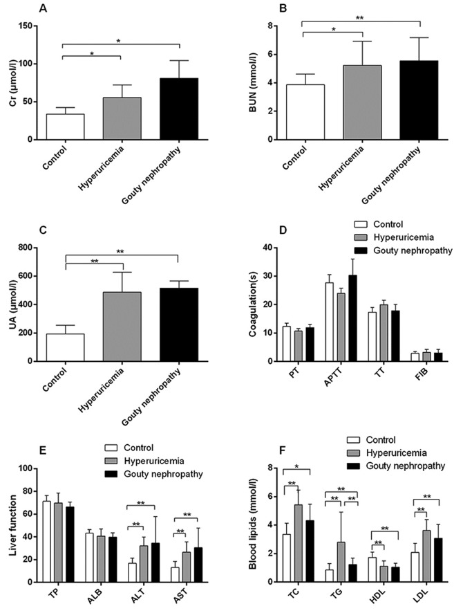
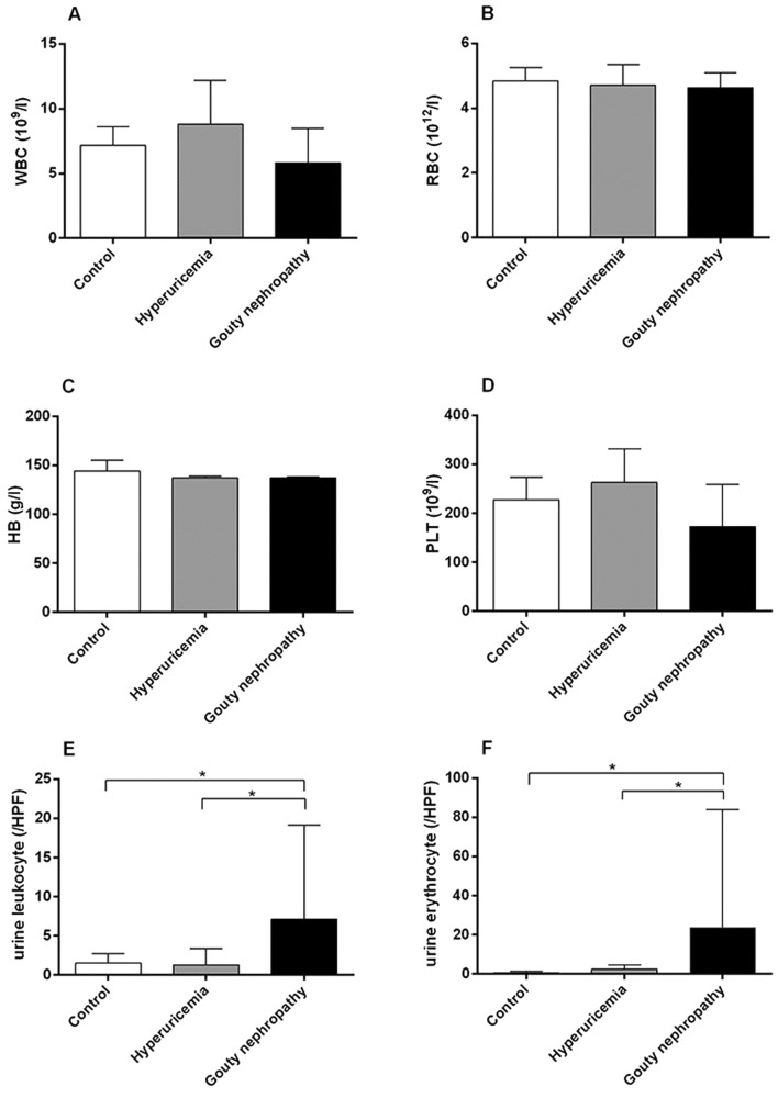
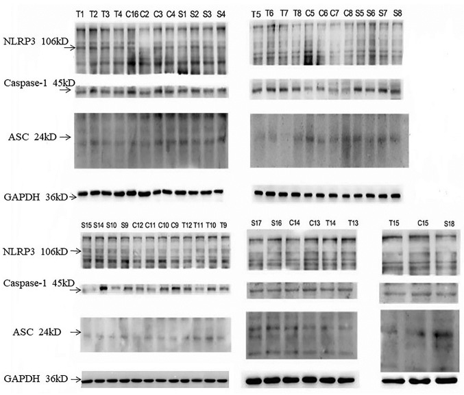
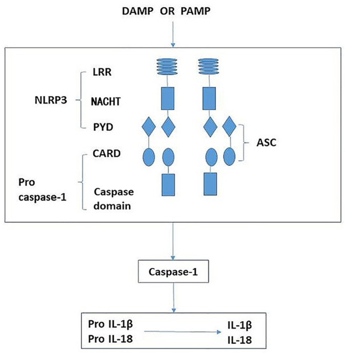
Crystalized deposits of monosodium urate activate the Nod-like receptor protein 3 (NLRP3) inflammasome, resulting in kidney damage. The present study investigated whether the NLRP3 inflammasome is associated with the progression of hyperuricaemia and gouty nephropathy. Adult male patients were recruited at the Affiliated Baoan Hospital of Shenzhen and divided into three groups of 15 patients each: The control group, the hyperuricaemia group and the gouty nephropathy group. General characteristics and organ function indicators were also measured for each patient. NLRP3, apoptosis-associated speck like protein (ASC) and caspase-1 mRNA and protein expressions in peripheral blood mononuclear cells were detected. The expression of certain downstream inflammatory factors, including interleukin (IL)-1β and IL-18 were also assessed in plasma. The results demonstrated that the concentration of uric acid and creatinine were increased in the hyperuricaemia and gouty nephropathy groups compared with the control group. NLRP3, ASC and caspase-1 mRNA and protein expression, and IL-1β and IL-18 expression were increased in the hyperuricaemia and gouty nephropathy groups compared with the control group. In addition, ASC and caspase-1 mRNA and protein expression, and IL-1β expression were higher in the gouty nephropathy group compared with the hyperuricaemia group. In conclusion, the present results supported the hypothesis that the NLRP3 inflammasome signalling pathway is associated with gouty nephropathy leading to initiation of the inflammatory response and causing renal damage.

Keywords: Nod-like receptor protein 3 inflammasome; gouty nephropathy; hyperuricaemia.


Figures

Figure 1
Figure 1
Organ function indicators of enrolled patients, including renal and liver function, coagulation and blood lipids. (A) Levels of Cr, (B) BUN, (C) UA, (D) coagulation functions, (E) liver function and (F) blood lipids of patients in the control, hyperuricemia and gouty nephropathy groups. *P<0.05 and **P<0.01, with comparisons indicated by lines. Cr, creatinine; BUN, blood urea nitrogen; UA, uric acid; PT, prothrombin time; APTT, activated partial thromboplastin time; TT, thrombin time; FIB, fibrinogen; TP, total protein; ALB, albumin; ALT, alanine aminotransferase; AST, aspartate aminotransferase; TC, total cholesterol; TG, total triglycerides; HDL, high density lipoprotein; LDL, low density lipoprotein.
Figure 2
Figure 2
Organ function indicators of enrolled patients, including blood and urine analysis. (A) WBC, (B) RBC, (C) HB, (D) PLT, (E) urine leukocytes and (F) urine erythrocytes levels of patients in the control, hyperuricemia and gouty nephropathy groups. *P<0.05, with comparisons indicated by lines. WBC, white blood cell; RBC, red blood cell; HB, hemoglobin; PLT, platelets; HPF, high power field.
Figure 3
Figure 3
Representative images of trypan blue staining of peripheral blood mononuclear cells (magnification, x10). Trypan Blue staining test was used to confirmed the cell viability, followed by the counting of cells. Trypan blue staining can stain dead cells blue, whilst the live cells are not colored. (A) Densely distributed colorless transparent particles under the microscope, representing live cells. (B) Scattered distribution of colorless transparent particles, showing fewer living cells. (C) Densely distributed colorless transparent particles, representing live cells. (D) Cell counting schematic. The area of every grid is 1.0 mm2 and the volume is 0.1 µl. Cells in the double-lined area were counted as red blood cells, whereas cells in the single-lined area counted as white blood cells. Magnification, x10.
Figure 4
Figure 4
Expression of NLRP3, ASC and caspase-1 protein in peripheral blood mononuclear cells. Representative western blots of peripheral blood mononuclear cells isolated from control, hyperuricemia and gouty nephropathy patients. NLRP3, Nod-like receptor protein 3; ASC, apoptosis-associated speck like protein; C, control group; T, hyperuricemia group; S, gouty nephropathy group.
Figure 5
Figure 5
Schematic demonstrating the activation of NLRP3 leading to release of proinflammatory factors. The NLRP3 inflammasome is comprised of NLRP3, ASC and caspase-1. When sensing danger signals, including PAMP and DAMP, NLRP3 oligomerizes via homotypic interactions between NACHT domains. The PYD of NLRP3 is then exposed for interaction with the PYD of ASC. The CARD of ASC in turn recruits pro-caspase-1 through CARD-CARD interactions, resulting in activated caspase-1. Activated caspase-1 then cleaves intracellular IL-1β and IL-18 precursors to form mature IL-1β and IL-18, which are secreted into the extracellular matrix. NLRP3, nucleotide-binding domain and leucine-rich repeat protein 3; ASC, apoptosis-associated speck like protein; LRR, leucine-rich repeat; NACHT, nucleotide-binding and oligomerization domain; PYD, pyrin domain; CARD, caspase recruitment domain; IL-1β, interleukin-1β; IL-18, interleukin-18; PAMP, pattern-associated molecular pattern; DAMP, danger-associated molecular pattern.

References

    1. Ryu ES, Kim MJ, Shin HS, Jang YH, Choi HS, Jo I, Johnson RJ, Kang DH. Uric acid-induced phenotypic transition of renal tubular cells as a novel mechanism of chronic kidney disease. Am J Physiol Renal Physiol. 2013;304(F471-F480) doi: 10.1152/ajprenal.00560.2012. - DOI - PubMed
    1. Siu YP, Leung KT, Tong MK, Kwan TH. Use of allopurinol in slowing the progression of renal disease through its ability to lower serum uric acid level. Am J Kidney Dis. 2006;47:51–59. doi: 10.1053/j.ajkd.2005.10.006. - DOI - PubMed
    1. Ghaemi-Oskouie F, Shi Y. The role of uric acid as an endogenous danger signal in immunity and inflammation. Curr Rheumatol Rep. 2011;13:160–166. doi: 10.1007/s11926-011-0162-1. [J] - DOI - PMC - PubMed
    1. Roddy E, Zhang W, Doherty M. The changing epidemiology of gout. Nat Clin Pract Rheumatol. 2007;3:443–449. doi: 10.1038/ncprheum0556. - DOI - PubMed
    1. Kim IY, Lee DW, Lee SB, Kwak IS. The role of uric acid in kidney fibrosis: Experimental evidences for the causal relationship. BioMed Res Int. 2014;2014(638732) doi: 10.1155/2014/638732. - DOI - PMC - PubMed