. 2020 Jun 2:11:2041731420926840.
doi: 10.1177/2041731420926840. eCollection 2020 Jan-Dec.

Osteogenic potential of poly(ethylene glycol)-amorphous calcium phosphate composites on human mesenchymal stem cells

Affiliations

Osteogenic potential of poly(ethylene glycol)-amorphous calcium phosphate composites on human mesenchymal stem cells

Aman S Chahal et al. J Tissue Eng. .

Abstract

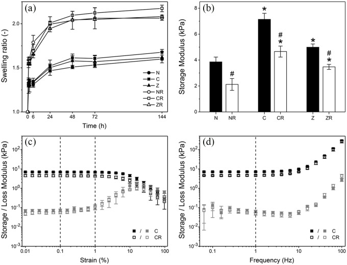
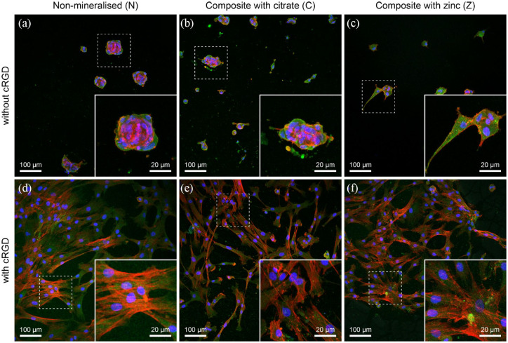
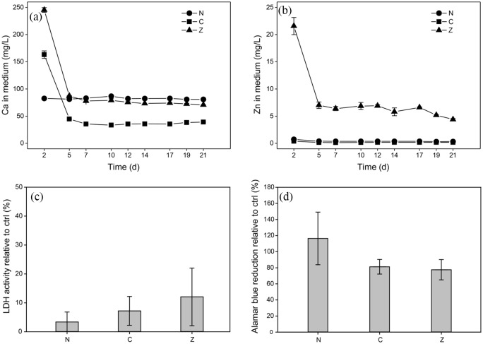
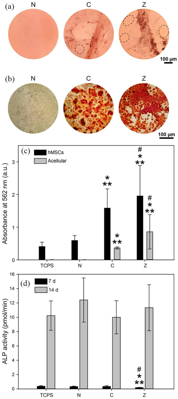
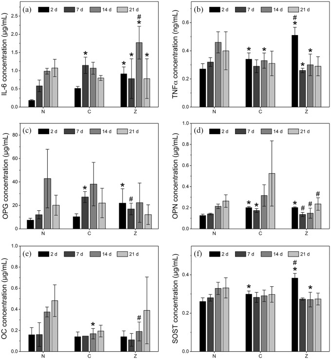
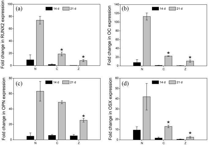
Synthetic hydrogel-amorphous calcium phosphate composites are promising candidates to substitute biologically sourced scaffolds for bone repair. While the hydrogel matrix serves as a template for stem cell colonisation, amorphous calcium phosphate s provide mechanical integrity with the potential to stimulate osteogenic differentiation. Here, we utilise composites of poly(ethylene glycol)-based hydrogels and differently stabilised amorphous calcium phosphate to investigate potential effects on attachment and osteogenic differentiation of human mesenchymal stem cells. We found that functionalisation with integrin binding motifs in the form of RGD tripeptide was necessary to allow adhesion of large numbers of cells in spread morphology. Slow dissolution of amorphous calcium phosphate mineral in the scaffolds over at least 21 days was observed, resulting in the release of calcium and zinc ions into the cell culture medium. While we qualitatively observed an increasingly mineralised extracellular matrix along with calcium deposition in the presence of amorphous calcium phosphate-loaded scaffolds, we did not observe significant changes in the expression of selected osteogenic markers.

Keywords: ACP; Hydrogel; PEG; calcium phosphate; cell differentiation; citrate; human mesenchymal stem cells; tissue engineering; zinc.


Conflict of interest statement

Declaration of conflicting interests: The author(s) declared no potential conflicts of interest with respect to the research, authorship, and/or publication of this article.

Figures

Figure 1.
Figure 1.
Mechanical characterisation of a non-mineralised hydrogel (N) and composites with citrate (C) or zinc (Z) depend on presence of mineral and cRGD-functionalisation (suffix R). (a) Volume swelling ratios versus time in cell culture medium. (b) Comparison of storage moduli of all sample groups as determined in oscillatory shear at 1 Hz and 0.1% strain. (c) Exemplary oscillatory shear strain sweeps of C composites with and without cRGD at 1 Hz frequency. (d) Exemplary frequency sweeps for the same samples at 0.1% strain. Mean values and standard deviation are shown for all samples (n = 3). * indicates significance compared to non-mineralised scaffolds with same cRGD-functionalisation, # indicates significance compared to cRGD-free scaffolds (p < 0.05).
Figure 2.
Figure 2.
Confocal images of hMSCs on hydrogels and composites with and without 2.5 mM of cRGD. hMSCs were immunolabelled for actin (red), vinculin (green) and DAPI (blue). (a-c) Representative images show cells seeded on non-mineralised (N) and composite hydrogels (C and Z) indicating the lack of spreading of hMSCs in the absence of cRGD. (d-f) Alternatively, images represent cells adopting a spread morphology when cultured on non-mineralised or composite hydrogels in the presence of cRGD tethered to the hydrogels.
Figure 3.
Figure 3.
(a, b) Calcium and zinc concentrations in cell culture media exhibit an initial burst release followed by steady concentration levels over time for both citrate- (C), and zinc-stabilised composites (Z). Calcium and zinc levels for non-mineralised hydrogels (N) correspond to the concentrations of the cell culture medium. Where error bars are not visible, standard deviations are smaller than symbols. Assessment of membrane integrity and cell metabolism of hMSCs after 72 h of exposure indicated no negative effects of mineralised and non-mineralised hydrogel scaffolds compared to TCPS controls. (c) Membrane integrity determined via extracellular LDH activity relative to controls (negative control, non-exposed cells on TCPS; positive control, cells lysed with Triton X-100). (d) Alamar blue reduction as a measure of cell metabolism relative to controls (negative control, non-exposed cells on TCPS; positive control, Alamar blue reagent in cell culture medium completely reduced by autoclaving). Graphs show mean values and standard deviations (n = 6).
Figure 4.
Figure 4.
(a) Phase contrast microscopy images reveal differences in cell sheet mineralisation of hMSCs after 21 d of exposure to non-mineralised (N), citrate- (C), and zinc-stabilised composites (Z). In addition, cell coverage varied considerably across the different groups (dashed ellipses indicate areas not covered by cells). (b) Alizarin red staining revealed increased ECM mineralisation for cells exposed to mineral-containing composites. (c) Quantification of alizarin red assay based on absorbance values at 562 nm (n = 4). (d) Membrane-bound ALP activity of hMSCs increased significantly between day 7 and 14 of exposure, indicating osteogenic differentiation irrespective of the scaffold group (n = 6). Bars represent mean values and standard deviation, **indicates statistical significance compared to TCPS control (cells not exposed to scaffold), *compared to N, and # compared to C (p < 0.05).
Figure 5.
Figure 5.
Cytokines and proteins measured from medium of hMSCs exposed to non-mineralised hydrogels (N), citrate- (C), and zinc-stabilised composites (Z). (a) Inflammation markers interleukin-6 (IL6) and (b) tumour necrosis factor α (TNFα) are shown among bone markers (c) osteoprotegerin (OPG), (d) osteopontin (OPN), (e) osteocalcin (OC), and (f) sclerostin (SOST). Osteogenic proteins such as OPG, OPN and OC are highest at day 14 or 21, indicating that cells are transitioning towards the osteogenic lineage. However, despite the initial release of calcium of zinc, no significant differences among the groups are observed at early timepoints. Bars represent mean values and standard deviations (n = 6). * indicates statistical significance compared to N, # compared to C (both p < 0.05).
Figure 6.
Figure 6.
RT-PCR of genes involved in osteogenic differentiation of hMSCs show a significant increase in gene expression between 14 and 21 d for (a) Runt-related transcription factor 2 (RUNX2), (b) Osteocalcin (OC), (c) Osteopontin (OPN), and (d) Osterix (OSX). Cells were cultured in the presence of non-mineralised hydrogels (N), citrate- (C), and zinc-stabilised composites (Z). Data is presented as fold-changes normalised to the housekeeping gene expression (GAPDH) levels. Bars represent the mean and standard deviation (n = 6), * indicates statistical significance compared to N.

References

    1. Giannoudis PV, Dinopoulos H, Tsiridis E. Bone substitutes: an update. Injury 2005; 36(Suppl. 3): 20–27. - PubMed
    1. Laurencin CT, El-Amin SF. Xenotransplantation in orthopaedic surgery. J Am Acad Orthop Surg 2008; 16(1): 4–8. - PubMed
    1. Arrington ED, Smith WJ, Chambers HG, et al. Complications of iliac crest bone graft harvesting. Clin Orthop Relat Res 1996; 329: 300–309. - PubMed
    1. Palmer SH, Gibbons CL, Athanasou NA. The pathology of bone allograft. J Bone Jt Surg Brit 1999; 81: 333–335. - PubMed
    1. Tomford WW. Transmission of disease through transplantation of musculoskeletal allografts. J Bone Joint Surg Am 1995; 77(11): 1742–1754. - PubMed

LinkOut - more resources