

Development of human prostate cancer stem cells involves epigenomic alteration and PI3K/AKT pathway activation
- PMID: 32537260
- PMCID: PMC7288500
- DOI: 10.1186/s40164-020-00168-0
Development of human prostate cancer stem cells involves epigenomic alteration and PI3K/AKT pathway activation
Abstract
Background: Human prostate cancer spheres endowed with stem cell properties have been obtained from androgen-dependent cell line LNCaP after exposure to an epigenomic modulator phenethyl isothiocynate (PEITC). Sphere cells can self-renew and grow with androgen, and also without androgen. Little is known about the signaling pathway and mechanism in the development of the stem cells in the spheres.
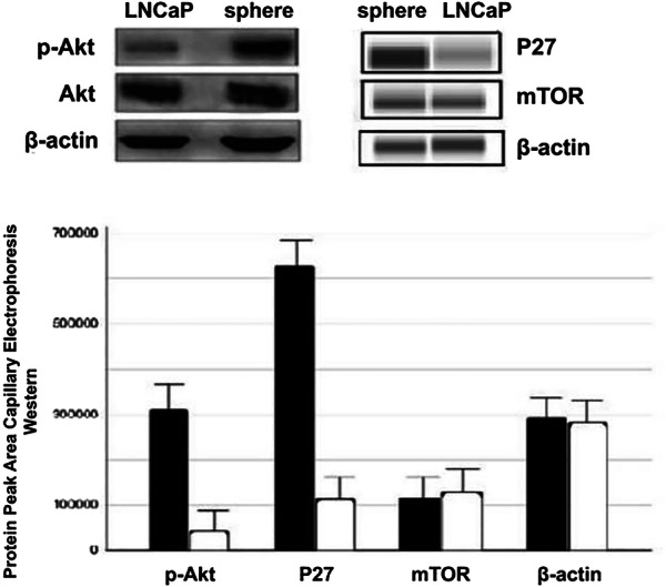
Methods: Expression of phosphoinositol-3 kinase (PI3K) pathway members and histone acetylation were quantified in the tumor spheres and LNCaP cells by western immunoblotting.
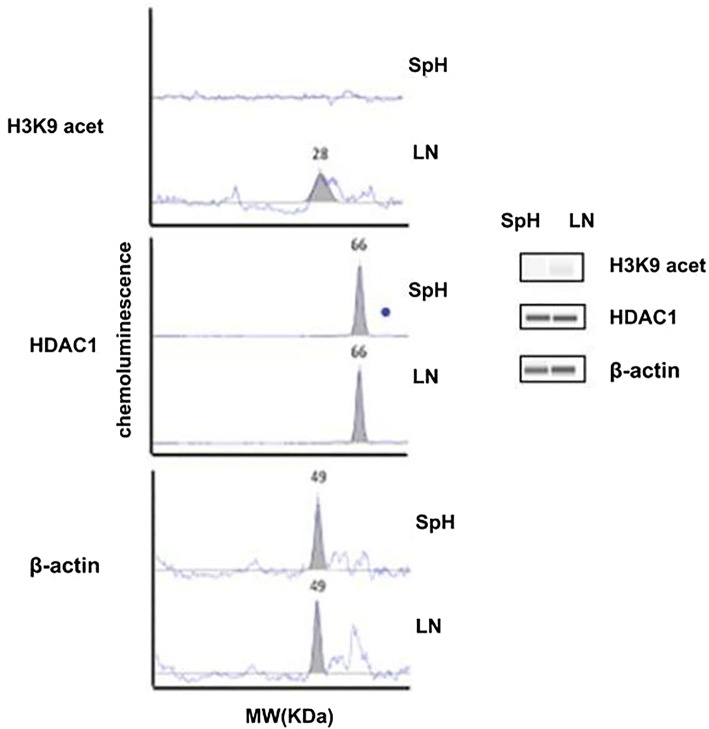
Results: The level of phosphorylated AKT was significantly increased in the sphere stem cells than the LNCaP cells at an average of 7.4 folds (range 5.8-10.7 folds), whereas the P27 level was elevated 5.4 folds (range 4.8-6.3 folds) (P < 0.05). The acetylation level on histone H3 lysine 9 was decreased.
Conclusions: PEITC appears to regulate the epigenome through histone acetylation and activate the PI3K/AKT pathway in the LNCaP cells. This mechanism may be responsible in part for the development of the prostate cancer stem cells.
Keywords: AKT; Cancer stem cells; Histone acetylation; PI3K; Prostate cancer; Sphere.
© The Author(s) 2020.
Conflict of interest statement
Competing interestsThe authors declare that they have no competing interests.
Figures
Similar articles
-
Establishment of prostate cancer spheres from a prostate cancer cell line after phenethyl isothiocyanate treatment and discovery of androgen-dependent reversible differentiation between sphere and neuroendocrine cells.Oncotarget. 2016 May 3;7(18):26567-79. doi: 10.18632/oncotarget.8440. Oncotarget. 2016. PMID: 27034170 Free PMC article.
-
Long-term androgen-ablation causes increased resistance to PI3K/Akt pathway inhibition in prostate cancer cells.Prostate. 2004 Feb 15;58(3):259-68. doi: 10.1002/pros.10332. Prostate. 2004. PMID: 14743465
-
Role of PI3K signaling in survival and progression of LNCaP prostate cancer cells to the androgen refractory state.Endocrinology. 2001 Nov;142(11):4795-805. doi: 10.1210/endo.142.11.8467. Endocrinology. 2001. PMID: 11606446
-
Reciprocal feedback inhibition of the androgen receptor and PI3K as a novel therapy for castrate-sensitive and -resistant prostate cancer.Oncotarget. 2015 Dec 8;6(39):41976-87. doi: 10.18632/oncotarget.5659. Oncotarget. 2015. PMID: 26506516 Free PMC article.
-
mTOR is a fine tuning molecule in CDK inhibitors-induced distinct cell death mechanisms via PI3K/AKT/mTOR signaling axis in prostate cancer cells.Apoptosis. 2016 Oct;21(10):1158-78. doi: 10.1007/s10495-016-1275-9. Apoptosis. 2016. PMID: 27484210
Cited by
-
Prostate cancer stem cells and their targeted therapies.Front Cell Dev Biol. 2024 Aug 8;12:1410102. doi: 10.3389/fcell.2024.1410102. eCollection 2024. Front Cell Dev Biol. 2024. PMID: 39175878 Free PMC article. Review.
-
The role of protein post-translational modifications in prostate cancer.PeerJ. 2024 Aug 12;12:e17768. doi: 10.7717/peerj.17768. eCollection 2024. PeerJ. 2024. PMID: 39148683 Free PMC article. Review.
-
The role of ncRNAs in neuroblastoma: mechanisms, biomarkers and therapeutic targets.Biomark Res. 2022 Apr 7;10(1):18. doi: 10.1186/s40364-022-00368-2. Biomark Res. 2022. PMID: 35392988 Free PMC article. Review.
-
Cellular mechanotransduction in health and diseases: from molecular mechanism to therapeutic targets.Signal Transduct Target Ther. 2023 Jul 31;8(1):282. doi: 10.1038/s41392-023-01501-9. Signal Transduct Target Ther. 2023. PMID: 37518181 Free PMC article. Review.
-
State-of-the-art therapeutic strategies for targeting cancer stem cells in prostate cancer.Front Oncol. 2023 Mar 9;13:1059441. doi: 10.3389/fonc.2023.1059441. eCollection 2023. Front Oncol. 2023. PMID: 36969009 Free PMC article. Review.
References
LinkOut - more resources
Full Text Sources
