. 2020 Jun 3;6(23):eaaz6486.
doi: 10.1126/sciadv.aaz6486. eCollection 2020 Jun.

Phase-dependent redox insulation in mussel adhesion

Affiliations

Phase-dependent redox insulation in mussel adhesion

Eric Valois et al. Sci Adv. .

Abstract

Catecholic 3,4-dihydroxyphenyl-l-alanine (Dopa) residues in mussel foot proteins (mfps) contribute critically to mussel (Mytilus californianus) plaque adhesion, but only if protected from oxidation at the adhesive-substratum interface. Dopa oxidation is thermodynamically favorable in seawater yet barely detectable in plaques; therefore, we investigated how plaques insulate Dopa-containing mfps against oxidation. Seawater sulfate triggers an mfp3 and mfp6 liquid-liquid phase separation (LLPS). By combining plaque cyclic voltammetry with electrophoresis, mass spectrometry, and redox-exchange chemistry, we show that Dopa-containing mfp3 and mfp6 in phase-separated droplets remain stable despite rapid oxidation in the surrounding equilibrium solution. The results suggest that a cohort of oxidation-prone proteins is endowed with phase-dependent redox stability. Moreover, in forming LLPS compartments, Dopa proteins become reservoirs of chemical energy.


Figures

Fig. 1
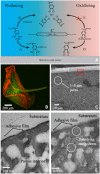
Fig. 1. Plaque chemistry and architecture.
(A) Dopa-containing mfps (spirals) recruited for adhesion by chemisorption (i) and cohesion by tris-catecholato Fe3+ complexation (ii). Dopa oxidized by O2 (iii) becomes dopaquinone, which forms covalent adducts but shows reduced surface binding (iv) and coordination (v). (B) Plaque with intrinsic fluorescence. (C) SEM of plaque section; (D) transmission electron microscopy of untreated plaque interface; (E) 5% acetic acid–treated (30 min) interface.
Fig. 2
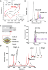
Fig. 2. Chemical analysis of plaque interfaces.
(A) CV plaque, pH 3 (5% acetic acid), and pH 8 (seawater) showing anodic peak potentials. Glassy carbon electrode at 50 mV/s scan rate. (B) XPS of plaque footprint on glass with sulfur speciation as thiol and sulfate peaks. a.u., arbitrary units. (C) Electroblotting strategy used to move soluble proteins: Plaque is positioned with adhesive interface facing a nitrocellulose membrane and the cathode. Nitrocellulose membrane was soaked in matrix, dried, and subjected to mass spectrometry. (D) Mass spectrum of electrokinetic proteins at pH 8 (red, tris glycinate) and pH 3 (black, acetic acid). Inset extends m/z range to 13,000. (E) Enlarged m/z of mfp3-1β showing mass register with mfp3-1β standard (purple).
Fig. 3
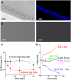
Fig. 3. Behavior of reconstituted plaque coacervates.
(A) Confocal images of settled (top) and (B) suspended (bottom) coacervate (Coa.) protein microdroplets. Visible micrograph (left) and fluorescent image (right). Scale bar: 5 μm. (C) Comparing redox exchange between 0.1 mM DPPH and plaque (plq) protein/sulfate coacervates or soluble (Sol.) plaque proteins. Oxidized DPPH is purple (λmax, 515 nm) and undergoes bleaching by reductive redox exchange with available Dopa and/or thiol groups. % change DPPH denotes [DPPH − DPPH2]/DPPH × 100 or the proportion by which initial DPPH color decreases over time. (D) Redox exchange between 10 mM periodate (Per) and soluble or coacervated plaque proteins at pH 3 and 8. At pH 8, oxidation occurs without periodate, and quinones add tris to increase in mass.
Fig. 4
Fig. 4. Phase-specific Dopa and thiol redox in coacervated plaque proteins.
(A) Schematic plaque interface. (B) Enlarged droplet with mfp3 and mfp6 variants. C1 is the film below the droplet and contains adhesive primers mfp5 and mfp8. When C1 Dopa is oxidized to dopaquinone (Ea + 660 mV Ag/AgCl; table S3), the damage is repaired with 2H+ + 2e donated by C2 (Ea + 540 mV). C2 dopaquinone is reduced back to Dopa by thiols in mfp6 (Ea, −200 mV), but when thiols are depleted, Dopa derivatives (∆-Dopa) provide reducing poise. Bidentate hydroquinone-quinone interactions actuate redox exchange in many cellular pathways (29).

References

    1. Hyman A. A., Weber C. A., Jülicher F., Liquid-liquid phase separation in biology. Annu. Rev. Cell Dev. Biol. 30, 39–58 (2014). - PubMed
    1. Vrhovski B., Jensen S., Weiss A. S., Coacervation characteristics of recombinant human tropoelastin. FEBS Journal 250, 92–98 (1997). - PubMed
    1. Jin H.-J., Kaplan D. L., Mechanism of silk processing in insects and spiders. Nature 424, 1057–1061 (2003). - PubMed
    1. Tan Y. P., Hoon S., Guerette P. A., Wei W., Ghadban A., Hao C., Miserez A., Waite J. H., Infiltration of a chitin by protein coacervates defines the squid beak mechanical gradient. Nat. Chem. Biol. 11, 489–495 (2015). - PubMed
    1. Yamanaka M., Ishizaki Y., Nagakawa T., Taoka A., Fukumori Y., Purification and characterization of coacervate-forming cuticular proteins from Papilio xuthus pupae. Zoolog. Sci. 30, 534–542 (2013). - PubMed

Publication types