EEG findings in acutely ill patients investigated for SARS-CoV-2/COVID-19: A small case series preliminary report
- PMID: 32537529
- PMCID: PMC7289172
- DOI: 10.1002/epi4.12399
EEG findings in acutely ill patients investigated for SARS-CoV-2/COVID-19: A small case series preliminary report
Erratum in
-
Erratum.Epilepsia Open. 2021 Jun;6(2):455. doi: 10.1002/epi4.12446. Epub 2020 Dec 25. Epilepsia Open. 2021. PMID: 34033705 Free PMC article. No abstract available.
Abstract
Objective: Acute encephalopathy may occur in COVID-19-infected patients. We investigated whether medically indicated EEGs performed in acutely ill patients under investigation (PUIs) for COVID-19 report epileptiform abnormalities and whether these are more prevalent in COVID-19 positive than negative patients.
Methods: In this retrospective case series, adult COVID-19 inpatient PUIs underwent EEGs for acute encephalopathy and/or seizure-like events. PUIs had 8-channel headband EEGs (Ceribell; 20 COVID-19 positive, 6 COVID-19 negative); 2 more COVID-19 patients had routine EEGs. Overall, 26 Ceribell EEGs, 4 routine and 7 continuous EEG studies were reviewed. EEGs were interpreted by board-certified clinical neurophysiologists (n = 16). EEG findings were correlated with demographic data, clinical presentation and history, and medication usage. Fisher's exact test was used.
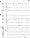
Results: We included 28 COVID-19 PUIs (30-83 years old), of whom 22 tested positive (63.6% males) and 6 tested negative (33.3% male). The most common indications for EEG, among COVID-19-positive vs COVID-19-negative patients, respectively, were new onset encephalopathy (68.2% vs 33.3%) and seizure-like events (14/22, 63.6%; 2/6, 33.3%), even among patients without prior history of seizures (11/17, 64.7%; 2/6, 33.3%). Sporadic epileptiform discharges (EDs) were present in 40.9% of COVID-19-positive and 16.7% of COVID-19-negative patients; frontal sharp waves were reported in 8/9 (88.9%) of COVID-19-positive patients with EDs and in 1/1 of COVID-19-negative patient with EDs. No electrographic seizures were captured, but 19/22 COVID-19-positive and 6/6 COVID-19-negative patients were given antiseizure medications and/or sedatives before the EEG.
Significance: This is the first preliminary report of EDs in the EEG of acutely ill COVID-19-positive patients with encephalopathy or suspected clinical seizures. EDs are relatively common in this cohort and typically appear as frontal sharp waves. Further studies are needed to confirm these findings and evaluate the potential direct or indirect effects of COVID-19 on activating epileptic activity.
Keywords: COVID‐19; SARS‐CoV‐2; encephalopathy; epileptiform discharges; seizures.
© 2020 The Authors. Epilepsia Open published by Wiley Periodicals LLC on behalf of International League Against Epilepsy.
Conflict of interest statement
AS Galanopoulou is co‐Editor in Chief of Epilepsia Open and has received royalties for publications from Elsevier and Morgan & Claypool publishers. AD Legatt serves on the editorial board of Journal of Clinical Neurophysiology and has received royalties for a publication from Springer Publishing. He has received consultant’s fees from Brain Sentinel. SR Haut serves on the editorial board of Epilepsy and Behavior. SL Moshé is serving as Associate Editor of Neurobiology of Disease and is on the editorial board of Brain and Development, Pediatric Neurology and Physiological Research. He receives from Elsevier an annual compensation for his work as Associate Editor in Neurobiology of Disease and royalties from two books he co‐edited. He has received consultant's fees from UCB and Pfizer. AB Boro is site PI for clinical trials sponsored clinical trials sponsored by Biogen, SK Life Science, Neurelis and UCB. He receives no salary support or other reimbursement for these projects. All funds go to the institution. None of the other authors have conflicts to disclose. We confirm that we have read the Journal's position on issues involved in ethical publication and affirm that this report is consistent with those guidelines. We confirm that we have read the Journal's position on issues involved in ethical publication and affirm that this report is consistent with those guidelines.
Figures

Comment in
-
COVID-19 EEG Studies: The Other Coronavirus Spikes We Need to Worry About.Epilepsy Curr. 2020 Sep 10;20(6):353-355. doi: 10.1177/1535759720956997. eCollection 2020 Nov-Dec. Epilepsy Curr. 2020. PMID: 34025253 Free PMC article. No abstract available.
References
Grants and funding
LinkOut - more resources
Full Text Sources
Miscellaneous

