. 2020 Jul;19(7):e13164.
doi: 10.1111/acel.13164. Epub 2020 Jun 14.

Geriatric fragility fractures are associated with a human skeletal stem cell defect

Affiliations

Geriatric fragility fractures are associated with a human skeletal stem cell defect

Thomas H Ambrosi et al. Aging Cell. 2020 Jul.

Abstract

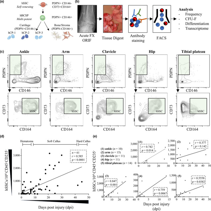
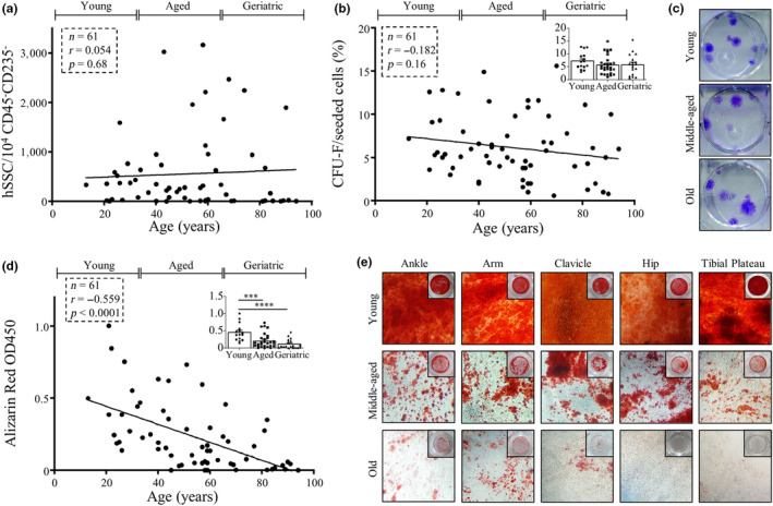
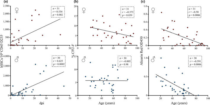
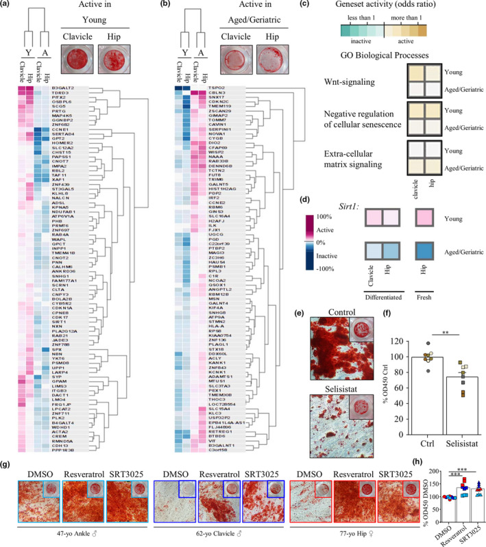
Fragility fractures have a limited capacity to regenerate, and impaired fracture healing is a leading cause of morbidity in the elderly. The recent identification of a highly purified bona fide human skeletal stem cell (hSSC) and its committed downstream progenitor cell populations provides an opportunity for understanding the mechanism of age-related compromised fracture healing from the stem cell perspective. In this study, we tested whether hSSCs isolated from geriatric fractures demonstrate intrinsic functional defects that drive impaired healing. Using flow cytometry, we analyzed and isolated hSSCs from callus tissue of five different skeletal sites (n = 61) of patients ranging from 13 to 94 years of age for functional and molecular studies. We observed that fracture-activated amplification of hSSC populations was comparable at all ages. However, functional analysis of isolated stem cells revealed that advanced age significantly correlated with reduced osteochondrogenic potential but was not associated with decreased in vitro clonogenicity. hSSCs derived from women displayed an exacerbated functional decline with age relative to those of aged men. Transcriptomic comparisons revealed downregulation of skeletogenic pathways such as WNT and upregulation of senescence-related pathways in young versus older hSSCs. Strikingly, loss of Sirtuin1 expression played a major role in hSSC dysfunction but re-activation by trans-resveratrol or a small molecule compound restored in vitro differentiation potential. These are the first findings that characterize age-related defects in purified hSSCs from geriatric fractures. Our results provide a foundation for future investigations into the mechanism and reversibility of skeletal stem cell aging in humans.

Keywords: aging; bone healing; geriatric fractures; human skeletal stem cell; sexual dimorphism.


Conflict of interest statement

The authors declare no competing interests.

Figures

Figure 1
Figure 1
Human SSCs accumulate upon fracture injury. (a) The human skeletal lineage tree. Human skeletal stem cells (hSSCs) sit at the apex of the lineage tree self‐renewing and giving rise to downstream cell populations including human bone, cartilage, stroma progenitors (hBCSP), human chondrogenic progenitors (hCP‐1, hCP‐2, and hCP‐3), bone, and stroma cell types. All cell populations are negative for CD45, CD235a, CD31, and Tie2. PDPN: podoplanin. (b) Experimental overview for the present study. (c) Representative FACS plots gated from singlet living cells gated negative for CD45, CD235a, CD31, and Tie2 showing the presence of hSSCs in callus tissue of fracture sites from various skeletal regions. Cells gated for PDPN+ and CD146 phenotype (upper panel) were further gated for CD73+ and CD164+ hSSCs (lower panel). (d) The correlation of hSSC prevalence and time (days post injury: dpi) is plotted for specimen used in this study. Demarcated healing stages are reflective of gross appearance of tissue at time of collection. (e) The same data are grouped and plotted by their origin of skeletal site. All graphs and data show two‐tailed Pearson correlation test results.
Figure 2
Figure 2
Geriatric fracture‐derived hSSC dysfunction occurs at the differentiation level. (a) The hSSC prevalence over donor age is plotted showing no correlation. (b) Fibroblast colony‐forming unit (CFU‐F) percentage is plotted over donor age. Top right, small plot: CFU‐F percentage was grouped by age into young (<35 years, n = 15), aged (35–65 years, n = 28), and geriatric (>65 years, n = 18). (c) Representative Crystal Violet staining for colonies formed by hSSCs derived from young, aged, and geriatric donors displaying similar quantities and sizes of colonies formed independent of age. (d) The correlation of osteogenic potential assessed by Alizarin Red S staining with donor age of fracture hSSCs is shown. Top right, small plot: Alizarin Red S staining measurement was grouped by age into young (<35 years, n = 15), aged (35–65 years, n = 28), and geriatric (>65 years, n = 18). (e) Representative Alizarin Red S stained culture images of differentiation outcome from young, aged, and geriatric donor fracture hSSCs from different skeletal sites are displayed. Linear correlations as analyzed with two‐tailed Pearson test. Grouped data plots are shown as mean + standard error of mean (SEM). Significance assessed by one‐way ANOVA (***p < .001 and ****p < .0001).
Figure 3
Figure 3
Female donor‐derived hSSCs display an exacerbated functional defect. (a) Correlation of hSSC prevalence at fracture site and days post injury (dpi) is shown (female: upper plot, male: lower plot). (b) Correlation of hSSC CFU‐F ability and donor age is shown (female: upper plot, male: lower plot). (c) Correlation of hSSC in vitro osteogenic potential and donor age is shown (female: upper plot, male: lower plot). Linear correlations as analyzed with two‐tailed Pearson test.
Figure 4
Figure 4
Transcriptomic differences of young and impaired aged hSSCs reveal Sirtuin1 as a potential target for hSSC rejuvenation. (a) Heatmap of microarray data showing the Top 80 genes uniquely active in young (29 and 26 years old) well‐differentiated (top panel: Alizarin Red S) or (b) age‐impaired (49 and 91 years old) ill‐differentiated (top panel: >45 years, Alizarin Red S) hSSCs from clavicle and hip fractures, respectively, as selected by highest dynamic range in dataset by GEXC. Hierarchical clustering of cell types and genes are shown. (c) Geneset activity of GO Biological Processes using GEXC showing either genesets uniquely active in young or aged/geriatric differentiated hSSCs. (d) Sirtuin1 gene expression as assessed by microarray in young hSSCs with strong osteogenic potential and aged hSSCs with impaired osteogenic potential derived from clavicle and hip fracture tissue (left). Sirt1 expression in freshly sorted young or aged hSSCs from hip (right). (e) Representative Alizarin Red S staining images of Sirt1 inhibition with Selisistat (1 µm) during osteogenic differentiation of hSSCs (from clavicle, arm and tibia fracture sites) at day 14. DMSO treated cells served as control. (f) Quantification of osteogenic potential by Alizarin Red S staining (n = 8, four different donors). (g) Representative Alizarin Red S staining images of three aged/geriatric patient‐derived hSSCs (two male and one female) with osteogenic differentiation defect (DMSO control) and treated with Sirtuin1 activators trans‐Resveratrol (2 µm) or small molecule SRT3025 (0.2 µm). (h) Quantification of osteogenic potential by Alizarin Red S staining in aged hSSCs from two male (blue) and one female (red) donors (n = 9, three different donors). Data shown as mean + standard error of mean (SEM). Significance assessed by one‐way ANOVA (***p < .001).

References

    1. Alswat, K. A. (2017). Gender disparities in osteoporosis. Journal of Clinical Medicine Research, 9, 382–387. 10.14740/jocmr2970w - DOI - PMC - PubMed
    1. Becker, D. J. , Kilgore, M. L. , & Morrisey, M. A. (2010). The societal burden of osteoporosis. Current Rheumatology Reports, 12, 186–191. 10.1007/s11926-010-0097-y - DOI - PubMed
    1. Caetano‐Lopes, J. , Lopes, A. , Rodrigues, A. , Fernandes, D. , Perpétuo, I. P. , Monjardino, T. , … Fonseca, J. E. (2011). Upregulation of inflammatory genes and downregulation of sclerostin gene expression are key elements in the early phase of fragility fracture healing. PLoS ONE, 6, e16947. - PMC - PubMed
    1. Carpintero, P. , Caeiro, J. R. , Carpintero, R. , Morales, A. , Silva, S. , & Mesa, M. (2014). Complications of hip fractures: A review. World Journal of Orthopedics, 5, 402–411. 10.5312/wjo.v5.i4.402 - DOI - PMC - PubMed
    1. Chan, C. K. F. , Gulati, G. S. , Sinha, R. , Tompkins, J. V. , Lopez, M. , Carter, A. C. , … Longaker, M. T. (2018). Identification of the human skeletal stem cell. Cell, 175, 43–56.e21. 10.1016/j.cell.2018.07.029 - DOI - PMC - PubMed

Publication types