Review
. 2020 Jul 21;49(14):5008-5057.
doi: 10.1039/c8cs01021h. Epub 2020 Jun 15.

Nanotechnology for angiogenesis: opportunities and challenges

Affiliations
Review

Nanotechnology for angiogenesis: opportunities and challenges

Saeid Kargozar et al. Chem Soc Rev. .

Abstract

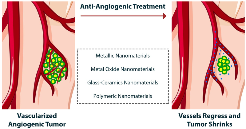
Angiogenesis plays a critical role within the human body, from the early stages of life (i.e., embryonic development) to life-threatening diseases (e.g., cancer, heart attack, stroke, wound healing). Many pharmaceutical companies have expended huge efforts on both stimulation and inhibition of angiogenesis. During the last decade, the nanotechnology revolution has made a great impact in medicine, and regulatory approvals are starting to be achieved for nanomedicines to treat a wide range of diseases. Angiogenesis therapies involve the inhibition of angiogenesis in oncology and ophthalmology, and stimulation of angiogenesis in wound healing and tissue engineering. This review aims to summarize nanotechnology-based strategies that have been explored in the broad area of angiogenesis. Lipid-based, carbon-based and polymeric nanoparticles, and a wide range of inorganic and metallic nanoparticles are covered in detail. Theranostic and imaging approaches can be facilitated by nanoparticles. Many preparations have been reported to have a bimodal effect where they stimulate angiogenesis at low dose and inhibit it at higher doses.


Figures

Fig. 1
Fig. 1
Schematic representation of different steps of angiogenic sprouting. A) The balance between pro-angiogenic signals (+) (e.g., VEGF), and anti-angiogenic factors (–) (e.g., tight pericyte (PC; yellow) contact), certain ECM molecules and VEGF inhibitors can control the sprouting. Under the appropriate conditions of angiogenesis, ECs can sprout (green), while others inhibit this phenomenon (grey). It has been well documented that the sprouting process needs to flip the apical-basal EC polarity, induce motile and invasive activity, modulate cell-cell contacts and degrade the local ECM. B) Attractive (+) or repulsive (–) cues from cells in the tissue environment are responsible for the growing EC sprouts. C) The fusion of adjacent sprouts into vessels occurs after adhesive or repulsive interactions between the cells at the tip. The fusion of vacuoles facilitates lumen formation in stalk ECs. D) A continuous lumen results from the fusion processes at the EC–EC interface; blood flow enhances oxygen delivery and subsequently reduces the hypoxia-induced pro-angiogenic signals. Maturation processes (e.g., the stabilization of cell junctions, matrix deposition, and tight PC attachment) is likely promoted by increased perfusion. Reproduced with permission from (Nature reviews Molecular cell biology, 2007, 8, 464–478), Copyright 2007, Nature Publishing Group.
Fig. 2
Fig. 2
Schematic illustration of (A) pro-angiogenic mediators and pathways involved in the activation of ECs and (B) the main clinical and preclinical factors involved in anti-angiogenic therapy.
Fig. 3
Fig. 3
The binding the IGF, an angiogenic molecule, to IGF-1R receptor on the cell surface activates two cell signaling pathways, leading to increased synthesis of HIF-1α by which the production of VEGF and thereby improved angiogenesis occur in the hypoxia condition.
Fig. 4
Fig. 4
Different types of NPs that have been used as therapeutics for anti-angiogenesis and vessel regression.
Fig. 5
Fig. 5
(A) Chemical structures of some of the most well-known pro- and anti-angiogenic substances derived from medicinal plants, and (B) the main signaling pathways of angiogenesis.
Fig. 6
Fig. 6
Pharmacokinetics and pharmacodynamics of resveratrol including bioavailability, anti-oxidant and inflammatory, anticancer, as well as healing properties, are enhanced when administered by nanocarriers in vivo. Reproduced with permission from (Colloids and Surfaces B: Biointerfaces, 2019, 180, 127–140), Copyright 2019, Elsevier Ltd.
Fig. 7
Fig. 7
Different molecular and cellular mechanisms of the antiangiogenic activity of paclitaxel. Reproduced with permission from (Angiogenesis, 2013, 16, 481–492), Copyright 2013, Springer Nature.
Fig. 8
Fig. 8
Schematic representation of the combined mechanism of CA4-NPs and sorafenib to treat hepatocellular carcinoma (HCC). As shown, although the disruption of established tumor blood vessels and extensive tumor necrosis are achieved by systemic administration of CA4-NPs, the overexpression of VEGF-A and thereby angiogenesis occurs in response to hypoxia. On the other hand, sorafenib can decrease the expression of VEGF-A and hence subsequently inhibit angiogenesis and tumor proliferation. This strategy could be considered as a potential approach to completely eradicate the whole tumor. Reproduced with permission from (Acta biomaterialia, 2019, 92, 229–240), Copyright 2019, Elsevier Ltd.
Fig. 9
Fig. 9
Schematic representation of the use of iRGD-PEI-MWNT-SS-CD/pAT2 for the inhibition of tumor angiogenesis. Intravenous administration of iRGD-PEI-MWNT-SS-CD/pAT2 complexes results in specific accumulation at tumor tissues via EPR effect; angiotensin II type 1 receptor (AT1R) and integrin receptor-mediated binding. Reproduced with permission from (Biomaterials, 2017, 139, 75–90), Copyright 2017, Elsevier Ltd.
Fig. 10
Fig. 10
Schematic representation of nanodiamond (ND)-induced vascular barrier leakiness. ND-induced vascular barrier leakiness leads to higher accumulation of doxorubicin in the tumor site. The increase of intracellular ROS and Ca2+ account for the ND-induced vascular barrier leakiness through the loss of cell-cell interconnection in the vascular barrier and cytoskeletal remodeling. Reproduced with permission from (ACS nano, 2016, 10, 1170–1181), Copyright 2016, American Chemical Society.
Fig. 11
Fig. 11
The images on the left side belong to anti-tumor metastasis activity of C60(OH)20; (A–C) Macroscopic observations of mice lungs soaked in Bouin’s solution exhibit spontaneous pulmonary breast cancer metastases (white arrows); (D–I) Pulmonary histology in mice received saline (D and G), 0.4 mg/kg (E and H) and 2 mg/kg C60(OH)20 (F and I). Note that black arrows indicate pulmonary metastases (original magnification: D–F ×100; G–I ×200). The images on the right side present the immunohistochemical staining of VEGF and CD31 expression to clarigy the effect of C60(OH)20 on EMT-6 tumor microvessel density; (A and D) Tumor tissues harvested from mice treated with saline; (B and E) 0.4 mg/kg C60(OH)20; and(C and F) 2 mg/kg C60(OH)20. Note that cells positive for VEGF and CD31 expression are in green and cell nuclei are blue (stained with DAPI). (×200 original magnification). Reproduced with permission from (Carbon, 2010, 48, 2231–2243), Copyright 2010, Elsevier Ltd.
Fig. 12
Fig. 12
Schematic illustration of the effect of environment on the pro-angiogenesis and anti-angiogenesis properties of nanoceria. The pH, reactive oxygen species (ROSs) generation, and intracellular oxygen concentration are identified as the main determinants of angiogenic behavior of nanoceria.
Fig. 13
Fig. 13
Representative schematic of cobalt roles in activating two signaling pathways involved in angiogenesis progress, i.e., the PI3K/Akt/mTOR and Ras/MEK/ERK pathway. It is assumed that cobalt via Akt activation could trigger transcription factors including SP1, NF-κB, RTEF and NFAT and thereby result in enhanced the transcription of HIF-1/2. Moreover, cobalt through activation of the MEK/ERK pathway could leads to the phosphorylation of 4E-BP and subsequent enhanced HIF-1 α translation.
Fig. 14
Fig. 14
Schematic representation of the angiogenesis regulation by copper ions. As seen, the entrance of copper into the cells is mediated by the copper transporter Ctr-1 and DMT1 proteins. Copper’s delivery to intracellular proteins is regulated by copper transport proteins (chaperones) such as Atox-1. By inhibiting PHD-mediated hydroxylation of HIF-1α, copper facilitates the translocation of the factor into the nucleus, leading to its dimerization with HIF-1 β and subsequent interactions to hypoxia-responsive elements and VEGF gene over-expression. Moreover, copper could activate the molecular signaling pathways resulting in increased NO and thereby promote angiogenesis.
Fig. 15
Fig. 15
Pro-angiogenic effect of copper nanoparticles: images of implants maintained in the chicken embryo chorioallantoic membrane (CAM) for 2 days, soaked with (1) control (non-soaked); (2) control (PBS); (3) CuSO4; (4) nano-copper, evaluated at day 12 of incubation. Scale bars, 2000 μm. Reproduced with permission from (International journal of molecular sciences, 2015, 16, 4838–4849), Copyright 2015, MDPI AG.
Fig. 16
Fig. 16
Redox signaling mechanism proposed for the pro-angiogenic effect induced by Eu(OH3) nanorods in endothelial cells (EC). ROSs (especially H2O2) are generated by the nanorods in the cytosolic part of the ECs, thus functioning as signaling molecules.
Fig. 17
Fig. 17
Overview of the size change of the large excision wounds made in the dorsal skin of diabetic mice with different periods (a) and relevant statistical analysis (b). Eu-MSNs-polymer film (Eu-P) significantly accelerated the wound healing compared to other groups. Masson’s Trichrome staining images (c) of wounds treated with different groups of films (blank control indicated as Ctrl, pure polymer film as Poly, MSNs-Polymer composite films as M-P and Eu-MSNs-Polymer composite films with as Eu-P). Green arrows indicates the newly formed epithelium at the wound site (scale bar 500 μm, *p < 0.05; **p < 0.01). Reproduced with permission from (Biomaterials, 2017, 144, 176–187), Copyright 2017, Elsevier Ltd.
Fig. 18
Fig. 18
Schematic illustration of molecular pathways affected by the anti-angiogenic effects of nano-gold (AuNPs). The main angiogenic pathways suppressed by nano-gold include VEGFR2, Tie2R, FGFR, and their downstream signaling pathways. As depicted, VEGF-165-mediated intracellular calcium release is suppressed by AuNPs. Moreover, AuNPs upregulate E-cadherin and downregulate vimentin, which results in reduced epithelial-to-mesenchymal transition (EMT) attenuating angiogenesis. Another anti-angiogenenic mechanism proposed for AuNPs is related to its ability to reduce ILs, MMPs, and TNF-α expression and inhibit neovascularization via induction of autophagy. Reproduced from (International Journal of Nanomedicine, 2019, 14, 7643), Copyright 2019, Dove Medical Press.
Fig. 19
Fig. 19
Schematic model of the molecular mechanisms on VEGFR2-mediated crosstalk between autophagy and angiogenesis signaling pathways triggered by silica nanoparticles (SiNPs).
Fig. 20
Fig. 20
Anti-angiogenic activity of Ag nanoparticles in vivo (rat model). Top panel: Gross photographs of Day 7 Matrigel implants with skin vessel background. Representative figures show (a) Streptozotocin without Ag nanoparticles, (b) Streptozotocin + Ag nanoparticles. Bottom panel: Histologic sections and hematoxylin and eosin-stained cross-sections showing representative photographs obtained from the sections of retina stained by hematoxylin and eosin in rats (c,d). Significant differences from control group were observed (p < 0.05). Reproduced with permission from (Biomaterials, 2009, 30, 6341–6350), Copyright 2009, Elsevier Ltd.
Fig. 21
Fig. 21
Schematic representation of the effect of AgNPs on angiogenesis process in cancer cells. AgNPs could enter the cells and prevent HIF-1α accumulation in the cytoplasm, followed by the suppression of HIF-1 target gene expression such as VEGF.
Fig. 22
Fig. 22
The possible molecular mechanisms and signaling pathways involved in zinc nanoflower-induced angiogenesis.
Fig. 23
Fig. 23
The use of NaLuF4: Yb,Tm@NaGdF4(153Sm) for four-modal imaging of tumor-bearing nude mice at 60 min post intravenous injection. A-D represent images obtained from upconversion luminescence (UCL), X-ray CT, SPECT, and MR of tumor, respectively. E exhibits UCL confocal image of the paraffin section of tumor tissue, and F is actually a schematic illustration of tumor angiogenesis imaging by applying the nanoparticles as the probe. Reproduced from (ACS Nano, 2013, 7, 11290–11300), Copyright 2013, American Chemical Society.

References

    1. Jain RK, Nature medicine, 2003, 9, 685. - PubMed
    1. Conway EM, Collen D and Carmeliet P, Cardiovascular research, 2001, 49, 507–521. - PubMed
    1. Carmeliet P and Jain RK, Nature, 2000, 407, 249–257. - PubMed
    1. Adams RH and Alitalo K, Nature reviews Molecular cell biology, 2007, 8, 464. - PubMed
    1. Schito L and Semenza GL, Trends in cancer, 2016, 2, 758–770. - PubMed