

Contrast enhanced ultrasound (CEUS) to assess pleural pulmonal changes in severe COVID-19 infection: First results
- PMID: 32538830
- PMCID: PMC7458509
- DOI: 10.3233/CH-209005
Contrast enhanced ultrasound (CEUS) to assess pleural pulmonal changes in severe COVID-19 infection: First results
Abstract
Aim: Use of contrast enhanced ultrasound (CEUS) in severe cases of COVID-19 infection to assess pulmonary changes near the pleura.
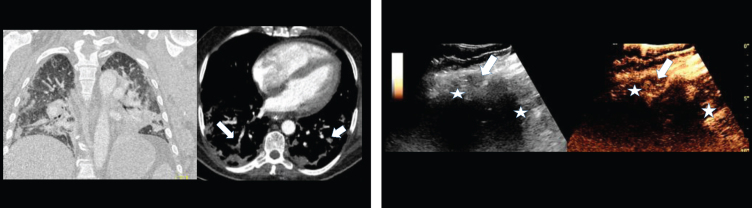
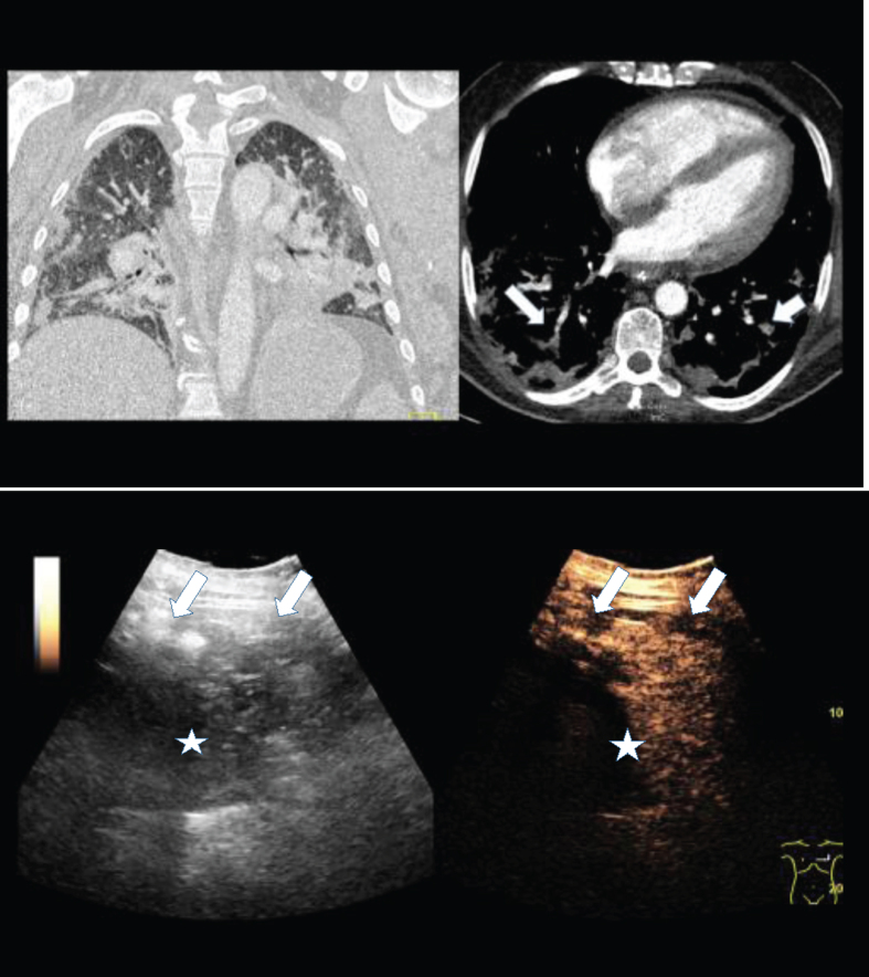
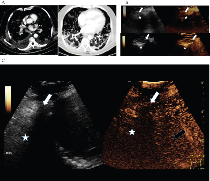
Material and methods: Bedside examinations by an experienced intensive care unit examiner using a multi-frequency probe (C1-6 MHz) with B-mode and CEUS to assess pleural-near changes in severe cases of COVID-19 infection with respiratory failure. CEUS with bolus delivery via a central venous catheter of 2.4 ml Sulphur hexafluoride microbubbles from the arterial phase (10-15 s) to the late phase of 5 min. Digital storage of cine sequences of the lung sound with abdomen for independent assessment with the subsequently performed contrast-enhanced dual-source CT.
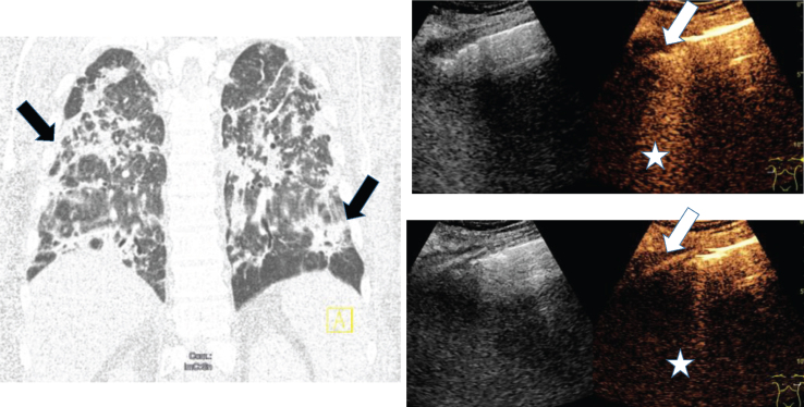
Results: In 11 intubated and ventilated patients (arithmetic mean 62 years, 48 to 78 years, 3 women) with confirmed severe COVID-19 infections, a peripherally accentuated consolidation with irregular hyperemia was found in the CEUS and also in the CT examination. Of the 5 cases with pulmonary arterial embolisms, signs of right ventricular failure were found. In all cases, using CEUS low perfused areas of the pleura with adjacent hyperemia could be detected, while, with CT segmental contrast medium, gaps with subpleural compressions were found. Interstitial changes near the pleura led to B-lines and to ground glass opacities in the CT. Near the diaphragm a delayed arterial contrast of the liver was observed. In addition, in 2 cases partial atelectasis, in 3 cases marginal pleural effusions were found.
Conclusion: CEUS opens up new possibilities for bedside monitoring of pleural reactive inflammatory or peripheral thrombus embolism in severe cases of COVID-19 infection.
Keywords: COVID-19 infection; lung- Contrast-enhanced ultrasound (CEUS); peripheral embolic consolidation; pleural affection.
Figures
Comment in
-
Complement-mediated Extracellular Vesicle release as a measure of endothelial dysfunction and prognostic marker for COVID-19 in peripheral blood - Letter to the Editor.Clin Hemorheol Microcirc. 2020;75(4):383-386. doi: 10.3233/CH-200958. Clin Hemorheol Microcirc. 2020. PMID: 32925002 No abstract available.
References
-
- Hu L, Wang C. Radiological role in the detection, diagnosis and monitoring for the coronavirus disease 2019 (COVID-19). Eur Rev Med Pharmacol Sci. 2020;24(8):4523–4528. - PubMed
-
- Dennie C, Hague C, Lim RS, Manos D, Memauri BF, Nguyen ET, Taylor J. Canadian Association of Thoracic Radiology/Canadian Association of Radiologists Consensus Statement Regarding Chest Imaging in Suspected and Confirmed COVID-19. Can Assoc Radiol J. 2020:846537120924606. - PubMed
MeSH terms
Substances
LinkOut - more resources
Full Text Sources
