. 2020 Aug 21;369(6506):956-963.
doi: 10.1126/science.abc7520. Epub 2020 Jun 15.

Isolation of potent SARS-CoV-2 neutralizing antibodies and protection from disease in a small animal model

Affiliations

Isolation of potent SARS-CoV-2 neutralizing antibodies and protection from disease in a small animal model

Thomas F Rogers et al. Science. .

Abstract

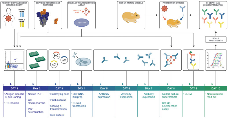
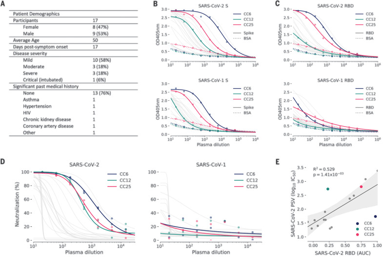
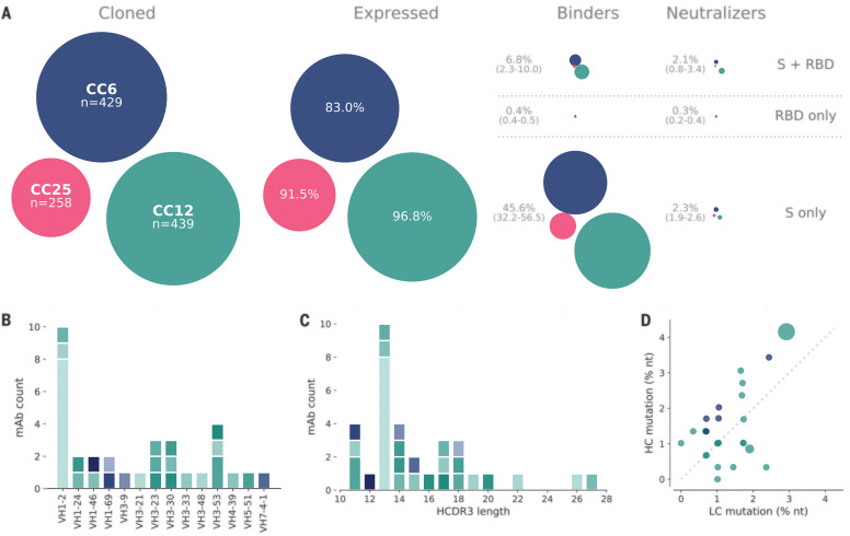
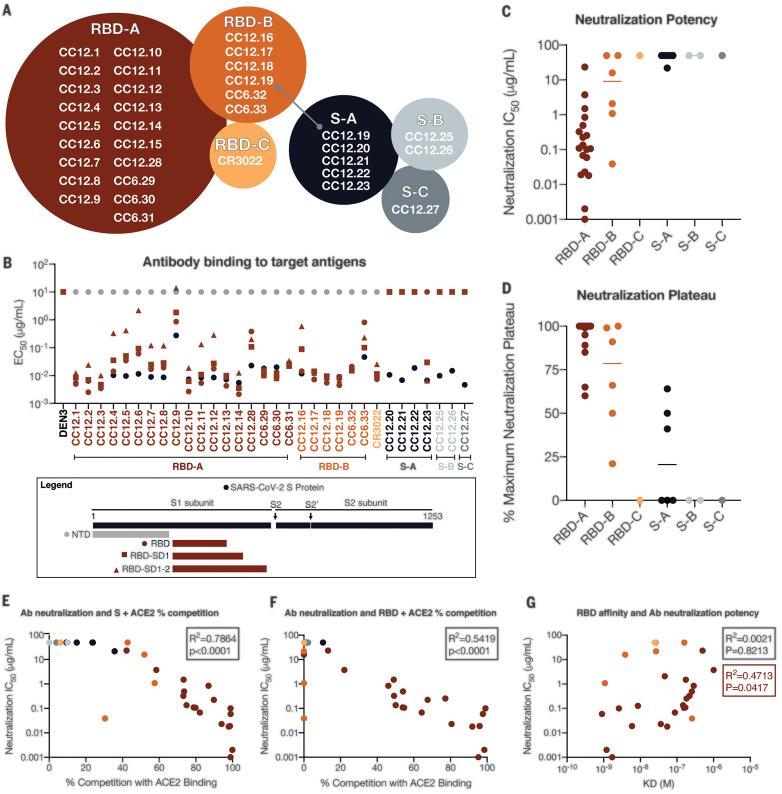
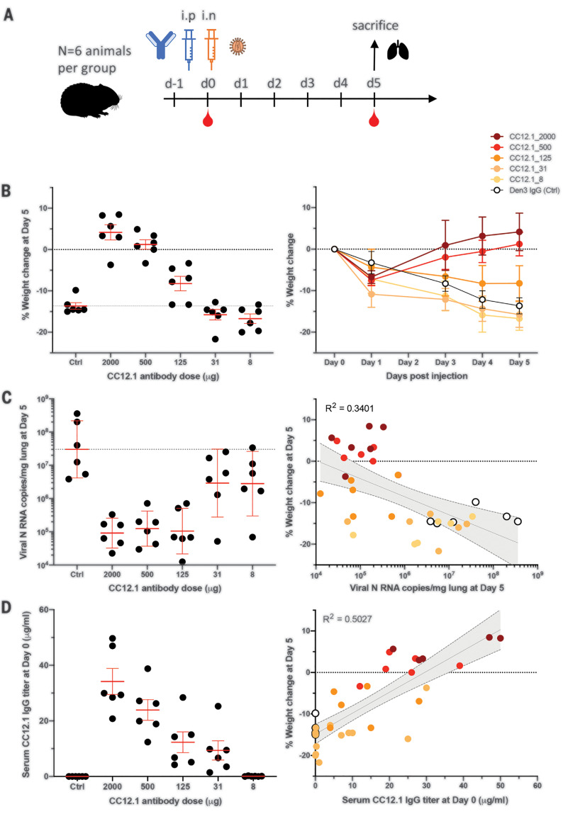
Countermeasures to prevent and treat coronavirus disease 2019 (COVID-19) are a global health priority. We enrolled a cohort of severe acute respiratory syndrome coronavirus 2 (SARS-CoV-2)-recovered participants, developed neutralization assays to investigate antibody responses, adapted our high-throughput antibody generation pipeline to rapidly screen more than 1800 antibodies, and established an animal model to test protection. We isolated potent neutralizing antibodies (nAbs) to two epitopes on the receptor binding domain (RBD) and to distinct non-RBD epitopes on the spike (S) protein. As indicated by maintained weight and low lung viral titers in treated animals, the passive transfer of a nAb provides protection against disease in high-dose SARS-CoV-2 challenge in Syrian hamsters. The study suggests a role for nAbs in prophylaxis, and potentially therapy, of COVID-19. The nAbs also define protective epitopes to guide vaccine design.


Figures

Fig. 1
Fig. 1. SARS-CoV-2 neutralizing antibody isolation strategy.
A natural infection cohort was established to collect plasma and PBMC samples from individuals who recovered from COVID-19. In parallel, functional assays were developed to rapidly screen plasma samples for SARS-CoV-2 neutralizing activity. SARS-CoV-2 recombinant surface proteins were also produced for use as baits in single-memory B cell sorting and downstream functional characterization of isolated mAbs. Finally, a Syrian hamster animal model was set up to evaluate mAb passive immunization and protection. The standard mAb isolation pipeline was optimized to facilitate high-throughput amplification, cloning, expression, and functional screening of hundreds of unpurified Ab heavy and light chain pairs isolated from each of several selected neutralizers in only 10 days. Selected pairs were scaled up to purify IgG for validation and characterization experiments. Potent neutralizing mAbs were selected to evaluate protection in the Syrian hamster model. HC, heavy chain; κC, kappa light chain; ΛC, lambda light chain; RT, reverse transcriptase.
Fig. 2
Fig. 2. COVID-19 cohort functional screening.
(A) Demographics of the University of California, San Diego (UCSD) COVID-19 cohort (CC) participants. CC plasma was tested for binding to SARS-CoV-1 and SARS-CoV-2 S proteins (B) and RBD subunits (C) by ELISA. Background binding of plasma to bovine serum albumin–coated plates is represented by a dotted line. OD405nm, optical density for wavelength of 405 nm. (D) Plasma was also tested for neutralization of pseudotyped (PSV) SARS-CoV-1 and SARS-CoV-2 virions. (E) Correlation between PSV SARS-CoV-2 neutralization and RBD subunit ELISA binding AUC (area under the curve). AUC was computed using Simpson’s rule. The 95% confidence interval of the regression line is shown by the gray shaded area and was estimated by performing 1000 bootstrap resamplings. R2 (coefficient of determination) and P values of the regression are also indicated. CC participants from whom mAbs were isolated are specifically highlighted in blue (CC6), green (CC12), and pink (CC25).
Fig. 3
Fig. 3. Antibody isolation and functional screening for SARS-CoV antigen binding and neutralization.
(A) Antibody down-selection process from three donors, presented as bubble plots. The areas of the bubbles for each donor are sized according to the number of antibodies (n) that were cloned and transfected, then scaled according to the number that were positive in subsequent assays. All antibodies that expressed at measurable levels were tested for binding to S protein and RBD to determine their specificity and then screened for neutralization. (B) VH gene distribution of down-selected mAbs. (C) Heavy chain CDR3 lengths of down-selected mAbs. Antibodies in (B) and (C) are colored according to their respective clonal lineages. (D) Mutation frequency of down-selected mAb lineages. Bubble position represents the mean mutation frequency for each lineage, with a bubble area that is proportional to lineage size. LC, light chain; nt, nucleotides.
Fig. 4
Fig. 4. Antibody functional activity by epitope specificities.
Monoclonal antibody epitope binning was completed using RBD and SARS-CoV-2 S protein as target antigens. (A) A total of three noncompeting epitopes for RBD (RBD-A, RBD-B, and RBD-C) and three noncompeting epitopes for S (S-A, S-B, and S-C) were identified. (B) MAbs were evaluated for binding to different target antigens (S, NTD, RBD, RBD-SD1, and RBD-SD1-2) by ELISA and apparent EC50 values are reported in micrograms per milliliter. (C) MAbs were evaluated for neutralization of SARS-CoV-2 pseudovirus using HeLa-ACE2 target cells. Antibodies are grouped according to epitope specificities, and neutralization IC50 values are reported in micrograms per milliliter. (D) The MNP is reported for each mAb and grouped by epitope specificity. MAbs were mixed with (E) S or (F) RBD protein and measured for binding to HeLa-ACE2 target cells as a measure of competition to the cell surface ACE-2 receptor. (G) mAb neutralization potencies (IC50) are plotted as a function of dissociation constants (KD) measured by SPR to RBD target antigen.
Fig. 5
Fig. 5. A potent SARS-CoV-2 RBD-specific neutralizing mAb protects against weight loss and lung viral replication in Syrian hamsters.
(A) SARS-CoV-2–specific human neutralizing mAb CC12.1 isolated from natural infection was administered at a starting dose of 2 mg per animal (average: 16.5 mg/kg) and subsequent serial fourfold dilutions. Control animals received 2 mg of Den3. Each group of six animals was challenged intranasally (i.n.) 12 hours after infusion with 1 × 10⁶ PFU of SARS-CoV-2. Serum was collected at the time of challenge (day 0), and animal weight was monitored as an indicator of disease progression. On day 5, lung tissue was collected for viral burden assessment. (B) Percent weight change was calculated from day 0 for all animals. (C) Viral load, as assessed by nucleocapsid RNA quantitative polymerase chain reaction (qPCR) from lung tissue at day 5 after infection. (D) Serum titers of the passively administered mAb, as assessed by ELISA at the time of challenge [12 hours after intraperitoneal (i.p.) administration]. Correlation analyses with 95% confidence intervals indicated by the gray shaded area.

Update of

Similar articles

Cited by

References

    1. The IMpact-RSV Study Group , Palivizumab, a humanized respiratory syncytial virus monoclonal antibody, reduces hospitalization from respiratory syncytial virus infection in high-risk infants. Pediatrics 102, 531–537 (1998). 10.1542/peds.102.3.531 - DOI - PubMed
    1. Corti D., Misasi J., Mulangu S., Stanley D. A., Kanekiyo M., Wollen S., Ploquin A., Doria-Rose N. A., Staupe R. P., Bailey M., Shi W., Choe M., Marcus H., Thompson E. A., Cagigi A., Silacci C., Fernandez-Rodriguez B., Perez L., Sallusto F., Vanzetta F., Agatic G., Cameroni E., Kisalu N., Gordon I., Ledgerwood J. E., Mascola J. R., Graham B. S., Muyembe-Tamfun J.-J., Trefry J. C., Lanzavecchia A., Sullivan N. J., Protective monotherapy against lethal Ebola virus infection by a potently neutralizing antibody. Science 351, 1339–1342 (2016). 10.1126/science.aad5224 - DOI - PubMed
    1. Misasi J., Gilman M. S. A., Kanekiyo M., Gui M., Cagigi A., Mulangu S., Corti D., Ledgerwood J. E., Lanzavecchia A., Cunningham J., Muyembe-Tamfun J. J., Baxa U., Graham B. S., Xiang Y., Sullivan N. J., McLellan J. S., Structural and molecular basis for Ebola virus neutralization by protective human antibodies. Science 351, 1343–1346 (2016). 10.1126/science.aad6117 - DOI - PMC - PubMed
    1. Walker L. M., Burton D. R., Passive immunotherapy of viral infections: ‘super-antibodies’ enter the fray. Nat. Rev. Immunol. 18, 297–308 (2018). 10.1038/nri.2017.148 - DOI - PMC - PubMed
    1. Giroglou T., Cinatl J. Jr.., Rabenau H., Drosten C., Schwalbe H., Doerr H. W., von Laer D., Retroviral vectors pseudotyped with severe acute respiratory syndrome coronavirus S protein. J. Virol. 78, 9007–9015 (2004). 10.1128/JVI.78.17.9007-9015.2004 - DOI - PMC - PubMed

Publication types

MeSH terms