. 2020 Jul 31;295(31):10511-10521.
doi: 10.1074/jbc.RA120.013976. Epub 2020 Jun 15.

The GTPase-activating protein p120RasGAP has an evolutionarily conserved "FLVR-unique" SH2 domain

Affiliations

The GTPase-activating protein p120RasGAP has an evolutionarily conserved "FLVR-unique" SH2 domain

Rachel Jaber Chehayeb et al. J Biol Chem. .

Abstract

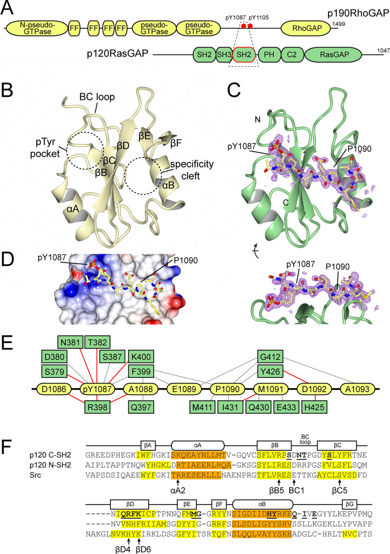
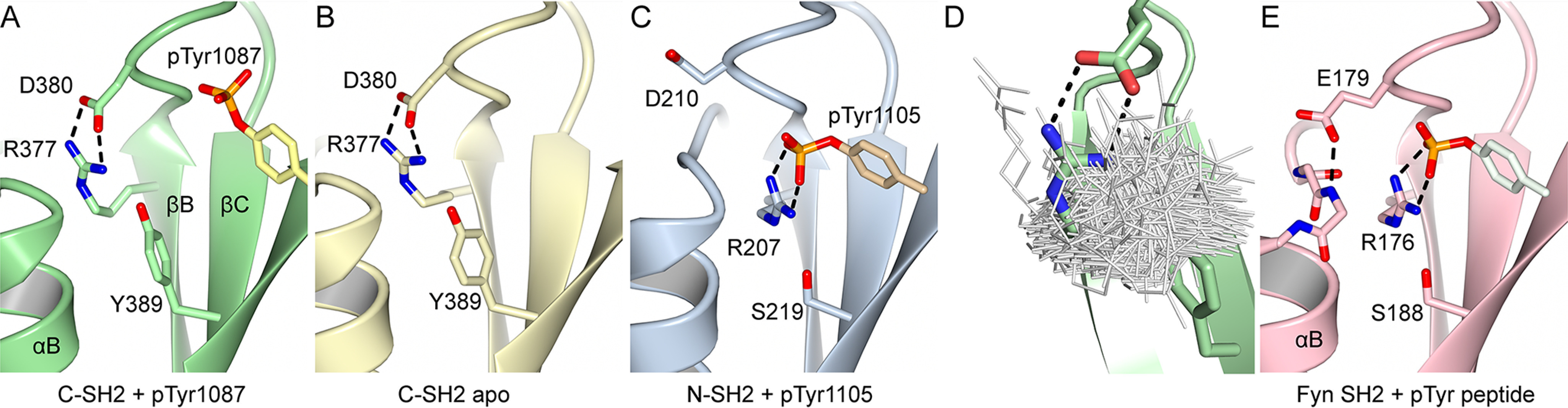
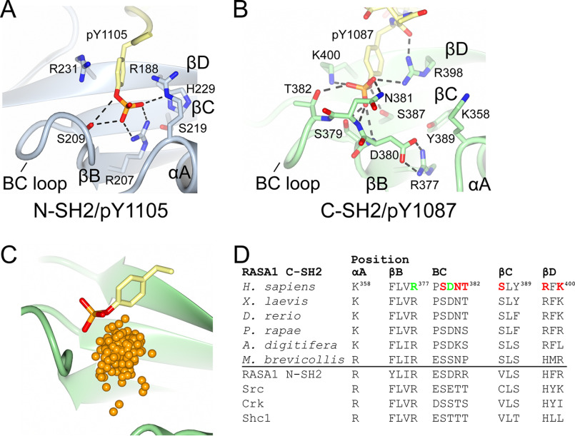
The Src homology 2 (SH2) domain has a highly conserved architecture that recognizes linear phosphotyrosine motifs and is present in a wide range of signaling pathways across different evolutionary taxa. A hallmark of SH2 domains is the arginine residue in the conserved FLVR motif that forms a direct salt bridge with bound phosphotyrosine. Here, we solve the X-ray crystal structures of the C-terminal SH2 domain of p120RasGAP (RASA1) in its apo and peptide-bound form. We find that the arginine residue in the FLVR motif does not directly contact pTyr1087 of a bound phosphopeptide derived from p190RhoGAP; rather, it makes an intramolecular salt bridge to an aspartic acid. Unexpectedly, coordination of phosphotyrosine is achieved by a modified binding pocket that appears early in evolution. Using isothermal titration calorimetry, we find that substitution of the FLVR arginine R377A does not cause a significant loss of phosphopeptide binding, but rather a tandem substitution of R398A (SH2 position βD4) and K400A (SH2 position βD6) is required to disrupt the binding. These results indicate a hitherto unrecognized diversity in SH2 domain interactions with phosphotyrosine and classify the C-terminal SH2 domain of p120RasGAP as "FLVR-unique."

Keywords: FLVR motif; GTPase-activating protein (GAP); RASA1; RasGAP; Src homology 2 domain (SH2 domain); X-ray crystallography; p120RasGAP; phosphotyrosine; protein structure; protein–protein interaction.


Conflict of interest statement

Conflict of interest—The authors declare that they have no conflicts of interest with the contents of this article.

Figures

Figure 1.
Figure 1.
Structure of p120RasGAP C-terminal SH2 domain. A, schematic of p120RasGAP interaction with p190RhoGAP. Domains are depicted, and the region co-crystallized is shown in a dashed box. B, structure of apo p120RasGAP C-SH2 indicating secondary structure elements and locations of the pTyr-binding pocket and specificity cleft. C, structure of p120RasGAP C-SH2 in complex with a synthesized phosphopeptide corresponding to p190RhoGAP-A residues 1086DpYAEPMDA1093. Simulated annealing omit FobsFcalc difference map contoured at 3σ RMSD is shown. D, surface electrostatics for p120RasGAP C-SH2 with p190RhoGAP-A peptide. E, interactions observed between p120RasGAP C-SH2 (green) and p190 phosphopeptide (yellow) defined by PDBsum (84). Hydrogen bonds are shown by red lines; nonbonded elements are shown by dashed lines. F, alignment of secondary structure features for p120RasGAP C-SH2, compared with p120RasGAP N-SH2 (PDB code 6PXC) (39), and Src (PDB code 1SPS) (18). β-Strands are highlighted yellow, and α-helices are highlighted orange. Residues that contact p190 phosphopeptide as defined by PDBsum are indicated with bold and underlined text. Residues discussed in the text are indicated.
Figure 2.
Figure 2.
An unusual conformation for the FLVR arginine of p120RasGAP C-SH2. A, crystal structure of phosphopeptide-bound p120RasGAP C-SH2 showing the FLVR arginine Arg377, Asp380, and Tyr389 and p190RhoGAP pTyr1087. B, crystal structure of apo p120RasGAP C-SH2. C, crystal structure of phosphopeptide-bound p120RasGAP N-SH2 (PDB code 2PXC) (39) showing the equivalent residues and phosphotyrosine. D, superposition of FLVR motif arginines from 592 Dali-aligned SH2 domains. E, crystal structure of Src family kinase Fyn (PDB code 4U1P) shows residue BC1 (Glu53) caps helix αB.
Figure 3.
Figure 3.
Phosphotyrosine-binding sites of p120RasGAP. A and B, detailed binding site interactions of the phosphotyrosine to p120RasGAP N-SH2 (PDB code 2PXC) (39) (A) and p120RasGAP C-SH2 (B). C, comparison of phosphotyrosine bound to p120RasGAP C-SH2 with the location of the phosphate atom in 245 phosphotyrosine-bound SH2 domains identified by the Dali server. Locations of the phosphates are shown as orange spheres. SH2 domains identified and superposed using by the Dali server (246 contain phosphotyrosine; 2BBU not included in analysis). D, conservation of key binding site residues over evolution and in human SH2 domains. pTyr-binding residues are colored red, and the FLVR arginine–Asp380 salt bridges are colored green.

References

    1. Liu B. A., Jablonowski K., Raina M., Arcé M., Pawson T., and Nash P. D. (2006) The human and mouse complement of SH2 domain proteins: establishing the boundaries of phosphotyrosine signaling. Mol. Cell 22, 851–868 10.1016/j.molcel.2006.06.001 - DOI - PubMed
    1. Sadowski I., Stone J. C., and Pawson T. (1986) A noncatalytic domain conserved among cytoplasmic protein-tyrosine kinases modifies the kinase function and transforming activity of Fujinami sarcoma virus P130gag-fps. Mol. Cell Biol. 6, 4396–4408 10.1128/mcb.6.12.4396 - DOI - PMC - PubMed
    1. Pawson T. (2004) Specificity in signal transduction: from phosphotyrosine–SH2 domain interactions to complex cellular systems. Cell 116, 191–203 10.1016/S0092-8674(03)01077-8 - DOI - PubMed
    1. Nash P. D. (2012) Why modules matter. FEBS Lett. 586, 2572–2574 10.1016/j.febslet.2012.04.049 - DOI - PubMed
    1. Shah N. H., Amacher J. F., Nocka L. M., and Kuriyan J. (2018) The Src module: an ancient scaffold in the evolution of cytoplasmic tyrosine kinases. Crit. Rev. Biochem. Mol. Biol. 53, 535–563 10.1080/10409238.2018.1495173 - DOI - PMC - PubMed

Publication types

MeSH terms

Substances

LinkOut - more resources