. 2020 Aug 20;64(9):e00623-20.
doi: 10.1128/AAC.00623-20. Print 2020 Aug 20.

Novel Aminoglycoside-Tolerant Phoenix Colony Variants of Pseudomonas aeruginosa

Affiliations

Novel Aminoglycoside-Tolerant Phoenix Colony Variants of Pseudomonas aeruginosa

Devin Sindeldecker et al. Antimicrob Agents Chemother. .

Abstract

Pseudomonas aeruginosa is an opportunistic bacterial pathogen and is known to produce biofilms. We previously showed the emergence of colony variants in the presence of tobramycin-loaded calcium sulfate beads. In this study, we characterized the variant colonies, which survived the antibiotic treatment, and identified three distinct phenotypes-classically resistant colonies, viable but nonculturable colonies (VBNC), and phoenix colonies. Phoenix colonies, described here for the first time, grow out of the zone of clearance of antibiotic-loaded beads from lawn biofilms while there are still very high concentrations of antibiotic present, suggesting an antibiotic-resistant phenotype. However, upon subculturing of these isolates, phoenix colonies return to wild-type levels of antibiotic susceptibility. Compared with the wild type, phoenix colonies are morphologically similar aside from a deficiency in green pigmentation. Phoenix colonies do not recapitulate the phenotype of any previously described mechanisms of resistance, tolerance, or persistence and, thus, form a novel group with their own phenotype. Growth under anaerobic conditions suggests that an alternative metabolism could lead to the formation of phoenix colonies. These findings suggest that phoenix colonies could emerge in response to antibiotic therapies and lead to recurrent or persistent infections, particularly within biofilms where microaerobic or anaerobic environments are present.

Keywords: Pseudomonas aeruginosa; aminoglycosides; antibiotic tolerance; biofilms.


Figures

FIG 1
FIG 1
In vitro imaging system (IVIS) images of P. aeruginosa Xen41 biofilm plates. (A) IVIS imaging was performed on day 0 of the tobramycin-loaded bead placement and daily thereafter until emergence of phoenix colonies was observed at day 4. Scale is at the right of the panel. Red indicates high levels of metabolic activity, and blue indicates low levels of metabolic activity. (B) IVIS imaging overlay on a black and white photo of the plate showing high levels of metabolic activity in all colonies at 5 days post-tobramycin-loaded bead placement.
FIG 2
FIG 2
Replica plating reveals resistant, phoenix colony, viable but nonculturable (VBNC), and persister cell variants. Images are of representative replica plates (n = 3). (A) Original plate before replica plating. (B) LB replica plate. (C) LB plus tobramycin replica plate. (A to C) Blue arrows represent colonies which grew on all three plate types and are thus resistant colonies. Green arrows represent colonies which appeared on the original and LB replica plate but not the LB replica plate containing tobramycin and are thus phoenix colonies. Purple arrows represent colonies which only grew on the original plate and thus are VBNCs. Yellow arrows represent likely persister cells, which did not appear on the original plate but grew on the LB replica plate only.
FIG 3
FIG 3
P. aeruginosa variant colonies emerge regardless of antibiotic delivery method. Colony counts were obtained of variants which emerged in response to tobramycin-loaded beads in each strain as well as to 1 mg of tobramycin in solution placed on a sterile filter disk in each strain. There was no significant difference in the number of phoenix colonies produced across all condition comparisons. Data are reported as the mean ± SD (n = 3).
FIG 4
FIG 4
Phoenix colonies have no defect in growth kinetics. (A) The OD600 was measured every 10 min for wild-type and phoenix isolates. Mean values (thick lines) and SD (thin lines) were plotted for resistant isolates (n = 7), phoenix colonies (n = 20), and wild-type P. aeruginosa Xen41 (n = 3). (B) Maximum specific growth rates were measured for each of the panel A curves. There was no significant difference between wild-type (WT) and resistant isolates (P = 0.3168) or between wild-type and phoenix colonies (P = 0.2981). (C) In situ colony growth was measured over time for resistant colonies (n = 29), phoenix colonies (n = 11), and VBNCs (n = 4) exposed to antibiotics. Time 0 represents the time at which the colonies were first noted. Although the phoenix colonies were consistently smaller, there was no significant difference found in colony size between any of the other groups at any time point. Data are reported as the mean ± SD.
FIG 5
FIG 5
Tobramycin concentration remains above the MIC during phoenix colony emergence. (A) A tobramycin-loaded bead was placed in sterile LB agar, and at various time points, agar plugs were extracted at various radii to examine the concentration of tobramycin by plating for MIC (n = 3) (data reanalyzed from reference 23). MIC zones were compared to a standard curve to calculate the tobramycin concentrations in the agar plugs. (B) At various radii, the lawn of a day 4 post-bead placement plate was resuspended, separated into fractions of freely diffusible and bound tobramycin, plated, and plotted against a standard curve to calculate the concentration of tobramycin. In the variant zone, both the intracellular and extracellular concentrations remain higher than the MIC. Data are reported as the mean ± SD.
FIG 6
FIG 6
Phoenix colony heritability is stable. (A) Variant colonies were generated as described, replica plated, and enumerated. Replica plating was performed, and counts were taken of the number of resistant and phoenix colonies. Phoenix colonies were isolated and grown overnight before being plated again. (B) Isolate cultures were then plated and allowed to grow for 5 days in the presence of a tobramycin-loaded bead. These plates were replica plated, and colony counts were obtained. Phoenix colonies were again isolated and grown in overnight broth cultures. (C) Isolate cultures from step B were plated and allowed to grow for 5 days in the presence of a tobramycin-loaded bead. Replica plating was again performed and colony counts obtained. No significant difference was seen in the number of phoenix colonies produced for each generation. Data are reported as the mean ± SD.
FIG 7
FIG 7
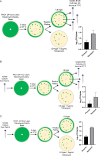
Anaerobic environments provide bacterial protection from antibiotics. Tobramycin-loaded beads were placed in the center of 24 h lawn biofilms grown on LB agar and LB agar containing 100 mM KNO3 in an anaerobic environment. The plates remained in the anaerobic environment for 5 days before being imaged using IVIS and replica plated on LB agar and LB agar containing 5 μg/ml of tobramycin. The replica plates were grown aerobically for 24 h before IVIS imaging was performed. While the anaerobic environment protected the lawn from killing, supplementing the plates with KNO3 allowed the lawn to be killed across most of the plate.
FIG 8
FIG 8
Antibiotic variant flow chart. Following the flow chart allows one to easily determine the antibiotic variant classification of cultures and isolates. Although this chart is based on a pregrown biofilm lawn of P. aeruginosa exposed to tobramycin, it can easily be adapted to other antibiotics, strains, and culturing techniques.

Similar articles

Cited by

References

    1. Shah NB, Osmon DR, Steckelberg JM, Sierra RJ, Walker RC, Tande AJ, Berbari EF. 2016. Pseudomonas prosthetic joint infections: a review of 102 episodes. J Bone Jt Infect 1:25–30. doi:10.7150/jbji.15722. - DOI - PMC - PubMed
    1. Serra R, Grande R, Butrico L, Rossi A, Settimio UF, Caroleo B, Amato B, Gallelli L, de Franciscis S. 2015. Chronic wound infections: the role of Pseudomonas aeruginosa and Staphylococcus aureus. Expert Rev Anti Infect Ther 13:605–613. doi:10.1586/14787210.2015.1023291. - DOI - PubMed
    1. Hoiby N, Ciofu O, Bjarnsholt T. 2010. Pseudomonas aeruginosa biofilms in cystic fibrosis. Future Microbiol 5:1663–1674. doi:10.2217/fmb.10.125. - DOI - PubMed
    1. Lewis K. 2008. Multidrug tolerance of biofilms and persister cells. Curr Top Microbiol Immunol 322:107–131. doi:10.1007/978-3-540-75418-3_6. - DOI - PubMed
    1. Zavascki AP, Carvalhaes CG, Picao RC, Gales AC. 2010. Multidrug-resistant Pseudomonas aeruginosa and Acinetobacter baumannii: resistance mechanisms and implications for therapy. Expert Rev Anti Infect Ther 8:71–93. doi:10.1586/eri.09.108. - DOI - PubMed

Publication types

LinkOut - more resources