. 2020 Jun 26;147(12):dev187344.
doi: 10.1242/dev.187344.

Differentiation of the human PAX7-positive myogenic precursors/satellite cell lineage in vitro

Affiliations

Differentiation of the human PAX7-positive myogenic precursors/satellite cell lineage in vitro

Ziad Al Tanoury et al. Development. .

Abstract

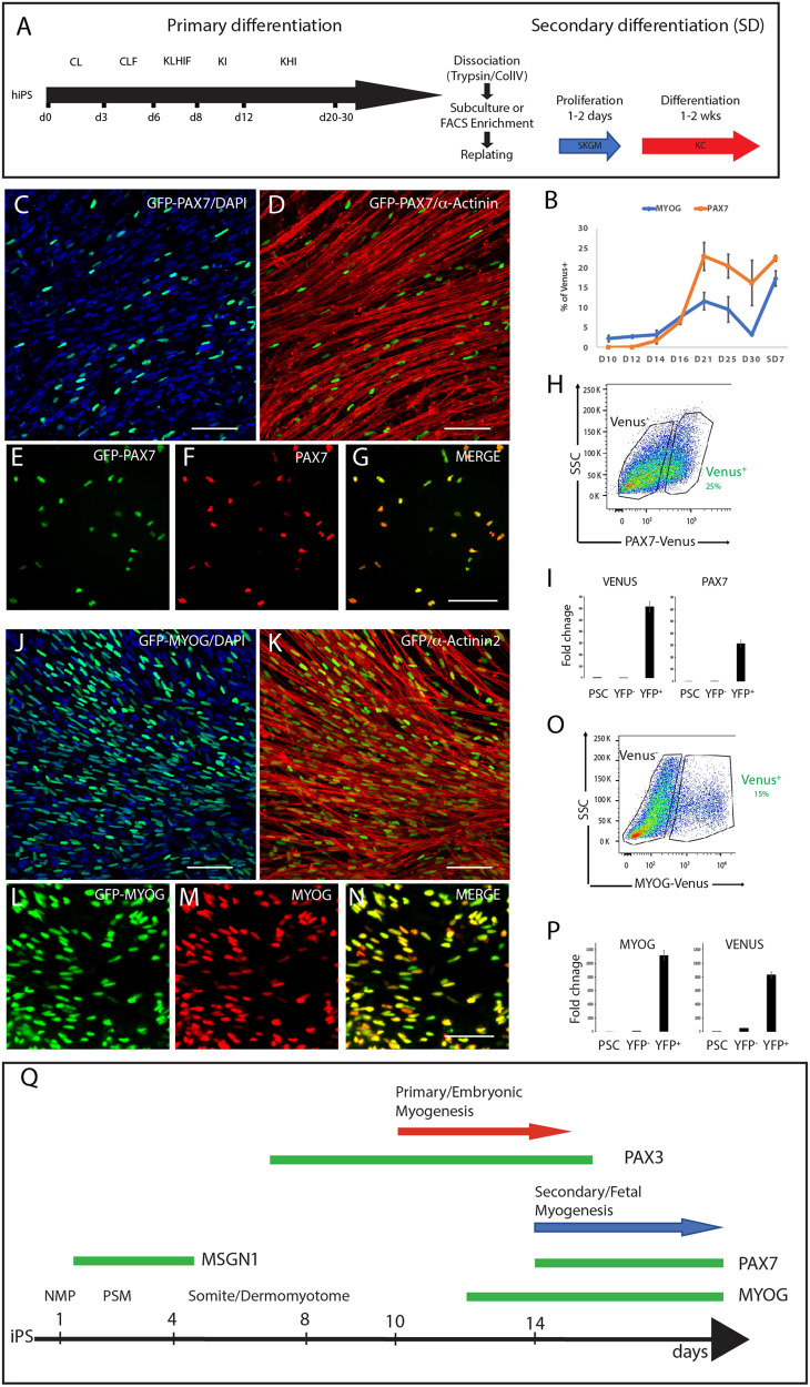
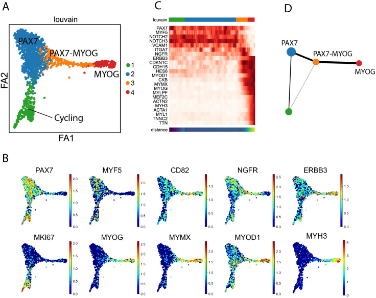
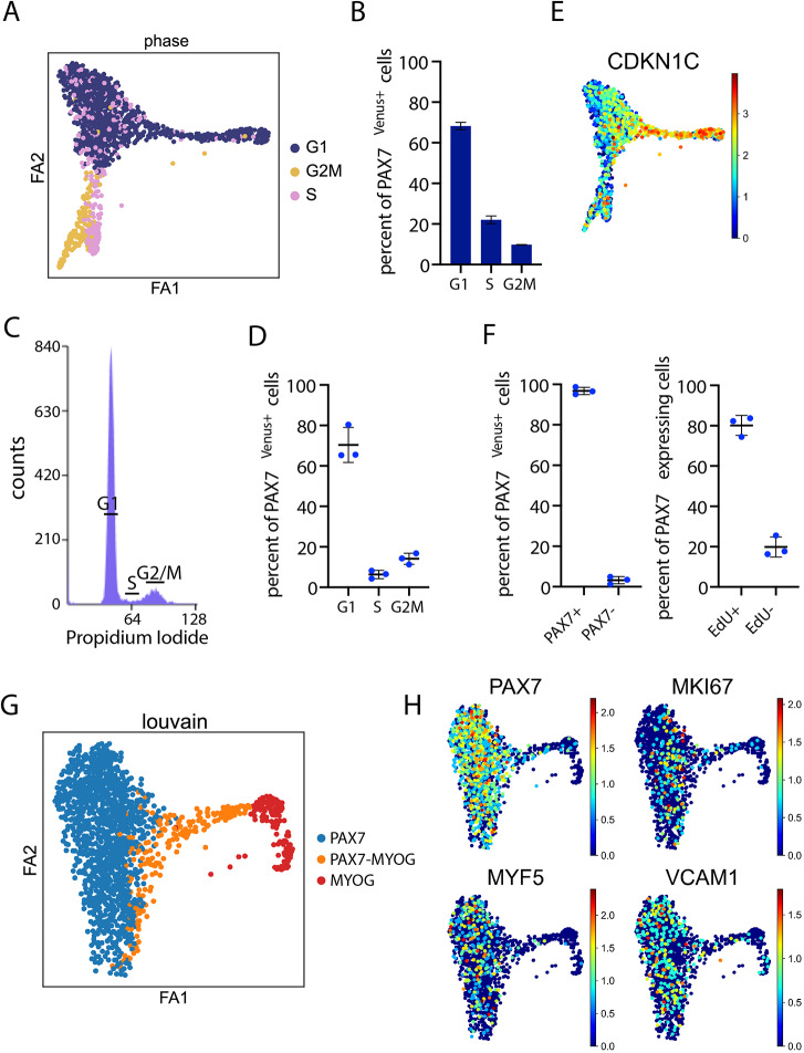
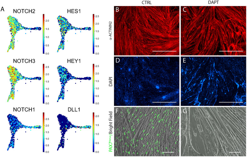
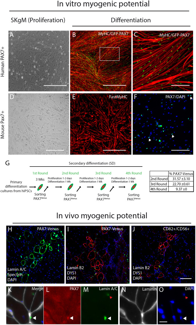
Satellite cells (SC) are muscle stem cells that can regenerate adult muscles upon injury. Most SC originate from PAX7+ myogenic precursors set aside during development. Although myogenesis has been studied in mouse and chicken embryos, little is known about human muscle development. Here, we report the generation of human induced pluripotent stem cell (iPSC) reporter lines in which fluorescent proteins have been introduced into the PAX7 and MYOG loci. We use single cell RNA sequencing to analyze the developmental trajectory of the iPSC-derived PAX7+ myogenic precursors. We show that the PAX7+ cells generated in culture can produce myofibers and self-renew in vitro and in vivo Together, we demonstrate that cells exhibiting characteristics of human fetal satellite cells can be produced in vitro from iPSC, opening interesting avenues for muscular dystrophy cell therapy. This work provides significant insights into the development of the human myogenic lineage.

Keywords: Human development; PAX7; Pluripotent stem cell; Satellite cell; Skeletal muscle.


Conflict of interest statement

Competing interestsOlivier Pourquie is a founder and shareholder of Anagenesis Biotechnologies.

Figures

Fig. 1.
Fig. 1.
Generation of human reporter iPSC lines. (A) Schematics of the differentiation protocol highlighting key factors and time scale of primary and secondary (following dissociation and replating) differentiation of myogenic progenitors from iPSC, as previously described (Chal et al., 2016). C, CHIR99021; d, day in culture; F, FGF-2; H, HGF; I, IGF; K, KSR; L, LDN193189. (B) FACS analysis showing the percentage of PAX7Venus+ and MYOGVenus+ cells in the mononucleated fraction during primary differentiation and after 7 days of secondary differentiation (SD7). (C-G) PAX7Venus reporter cells at SD7 stained with antibodies against GFP and α-actinin (C,D) or with anti-GFP and anti-PAX7 antibodies (E-G). (H) Flow cytometry analysis of PAX7Venus cultures. PAX7Venus reporter cells were differentiated as described above for primary differentiation. Cultures were dissociated after 3 weeks and FACS sorted. (I) RT-qPCR analysis for Venus and PAX7 in undifferentiated human iPSC (PSC) and PAX7Venus+ FACS-sorted cells from 3-week-old primary cultures. (J-N) MYOGVenus reporter cultures at SD7 stained with antibodies against GFP and α-actinin (J,K) or with anti-GFP and anti-MYOG antibodies (L-N). Nuclei were counterstained with DAPI. (O) Flow cytometry analysis of MYOGVenus cultures. MYOGVenus reporter cells were differentiated for 2 weeks in primary differentiation, dissociated and FACS sorted. (P) RT-qPCR analysis for Venus and MYOG in undifferentiated human iPSC and FACS-sorted MYOGVenus+ cells isolated from 3-week-old primary cultures. (Q) Schematic recapitulating the differentiation timeline of human iPSC differentiating to skeletal muscle in vitro. Green bars represent expression windows of the fluorescent reporter lines. NMP, neuro-mesodermal precursors; PSM, presomitic mesoderm. Scale bars: 100 µm.
Fig. 2.
Fig. 2.
Single cell analysis of PAX7Venus+ cells. (A) ForceAtlas2 force-directed layout (k=10, 30 PC dimensions, 1427 cells) of single cell transcriptomes from 30-day-old FACS-sorted PAX7Venus+ cells from primary cultures. Colors indicate Louvain cluster IDs. (B) Genes indicative of cluster 2 (PAX7, MYF5), cluster 1 (MKI67), cluster 3 (MYOG, MYMX) and cluster 4 (MYH3), as well as markers of human myogenic precursors (MYOD1, CD82, NGFR, ERBB3) shown in ForceAtlas2 layouts colored by log-normalized transcript counts. (C) Pseudo-temporal ordering of PAX7Venus+ cells along a path towards a differentiated state. Top bar, colors indicate Louvain IDs. Bottom, heatmap of selected markers showing gene changes along PAGA path (root=cluster 1). (D) Coarse-grained layout of PAX7Venus+ cells using Partition-based graph abstraction (PAGA).
Fig. 3.
Fig. 3.
Cell cycle analysis of PAX7Venus+ cells. (A,B) Cell cycle analysis of the PAX7Venus+ fraction by scRNA-seq. PCA plot showing distribution of cell cycle states in PAX7Venus+ cells identified based on scRNA-seq (A). Quantification of cells in different cell cycle states (1427 cells, mean±s.d., n=2) (B). (C,D) Cell cycle analysis of the PAX7Venus+ fraction by DNA content analysis using propidium iodide. (C) Representative frequency histogram showing distribution of cells with different DNA content. (D) Quantification of PAX7Venus+ cells in different cell cycle phases using DNA propidium iodide staining. (E) Force Atlas2 layouts colored by log-normalized transcript counts of CDKN1C. (F) Percent of PAX7Venus+ cells expressing the PAX7 protein (left). Quantification of PAX7 protein expressing cells labeled by EdU after 40 h (right) (mean±s.d., n=3). (G) ForceAtlas2 layout and Louvain clustering after performing the regression of cell cycle genes. Colors indicate Louvain cluster IDs. (H) After cell cycle regression cycling cells collapse with cluster 1 (MYF5). Cell cycle indicator MKI67 and other genes shown in ForceAtlas2 layouts colored by log-normalized transcript counts.
Fig. 4.
Fig. 4.
Notch signaling is required for the maintenance of the PAX7Venus+ cells in vitro. (A) Distribution and expression of the Notch receptors (NOTCH1, NOTCH2 and NOTCH3), the ligand DLL1, and the targets (HEY1 and HES1) in the PAX7+/MYOG clusters. (B-G) Effect of Notch signaling on the differentiation of human PAX7 precursors in vitro. PAX7Venus+ cells were FACS-sorted after 3 weeks of primary differentiation, replated as described and differentiated for 1 week in KC differentiation medium in the absence or presence of the γ-secretase inhibitor DAPT (25 µM) that blocks Notch signaling. Cells were fixed and stained with α-actinin and GFP antibodies. Scale bars: 1000 µm in B-E; 100 µm in F,G.
Fig. 5.
Fig. 5.
Myogenic potential of iPSC-derived PAX7-Venus. (A-F) Myogenic potential of purified human (A-C) PAX7Venus+ and mouse (D-F) Pax7GFP+ cells in vitro. Human iPSC and mouse ESC were differentiated for 3 weeks as previously described (Chal et al., 2016) and fluorescent cells were FACS-isolated and replated in SkGM medium for 1-2 days (A,D) to allow 80-90% confluency. Cells were then induced for differentiation and immunostained for GFP and FAST MyHC (B,C,E,F). Nuclei were counterstained with DAPI. (G) Serial sorting and differentiation of human PAX7Venus+ cells. PAX7Venus+ cells were differentiated as described for primary differentiation, dissociated at 3 weeks and FACS sorted (first round). FACS-sorted cells were then replated in SkGM medium for 1-2 days to allow 80-90% confluency and then differentiated for 1 week in a differentiation medium containing 1 µm CHIR99021 and 2% KSR. Cultures were then dissociated and FACS-sorted for Venus expression (second round). Replating and sorting experiments were repeated as indicated. The percentage of PAX7Venus+ cells is shown at each round (n=3). (H-O) Transplantation of human PAX7Venus+ cells in immune-deficient mice: 105 FACS-sorted PAX7Venus+ cells were injected into the TA of cardiotoxin-injured Rag2B6 WT (H) or NOD-Rag1−/− Dmdmdx-5cv mice (I,J). In vivo contribution of the transplanted PAX7Venus+ human cells to muscle fibers is visualized on transverse sections of the grafted TA muscle. Sections were stained with a combination of human-specific anti-spectrin (green)/laminA/C (green) (H), which detect human fibers and human nuclei, respectively, but not mouse myofibers, and with anti-dystrophin (red) and lamin B2 (green) antibodies (I) that co-detect dystrophin+ myofibers and human-derived nuclei, respectively. J shows a transverse section of TA muscle grafted with FACS-sorted CD82+/CD56+ primary fetal human myogenic precursor cells and stained with anti-dystrophin and anti-human lamin B2. (K-O) Higher magnification of grafted TA section showing a human PAX7Venus+ cell (white arrowhead) expressing the PAX7 protein (L) and human specific lamin A/C (M) localized under the laminin+ basal lamina (N) (chicken anti-laminin antibody). Nuclei are labeled with DAPI (O). Red arrow shows a human laminA/C+ PAX7 myofiber nucleus. Scale bars: 200 µm in A,D,C,F; 1000 µm in B,E; 100 µm in H-J; 50 µm in K-O.

References

    1. Alexander M. S., Rozkalne A., Colletta A., Spinazzola J. M., Johnson S., Rahimov F., Meng H., Lawlor M. W., Estrella E., Kunkel L. M. et al. (2016). CD82 is a marker for prospective isolation of human muscle satellite cells and is linked to muscular dystrophies. Cell Stem Cell 19, 800-807. 10.1016/j.stem.2016.08.006 - DOI - PMC - PubMed
    1. Barruet E., Garcia S. M., Striedinger K., Wu J., Lee S., Byrnes L., Wong A., Xuefeng S., Tamaki S., Brack A. S. et al. (2020). Functionally heterogeneous human satellite cells identified by single cell RNA sequencing. eLife 9, e51576 10.7554/elife.51576 - DOI - PMC - PubMed
    1. Becht E., McInnes L., Healy J., Dutertre C.-A., Kwok I. W. H., Ng L. G., Ginhoux F. and Newell E. W. (2018). Dimensionality reduction for visualizing single-cell data using UMAP. Nat. Biotechnol. 37, 38-44. 10.1038/nbt.4314 - DOI - PubMed
    1. Belle M., Godefroy D., Couly G., Malone S. A., Collier F., Giacobini P. and Chédotal A. (2017). Tridimensional visualization and analysis of early human development. Cell 169, 161-173.e12. 10.1016/j.cell.2017.03.008 - DOI - PubMed
    1. Bi P., Ramirez-Martinez A., Li H., Cannavino J., McAnally J. R., Shelton J. M., Sánchez-Ortiz E., Bassel-Duby R. and Olson E. N. (2017). Control of muscle formation by the fusogenic micropeptide myomixer. Science 356, 323-327. 10.1126/science.aam9361 - DOI - PMC - PubMed

Publication types

MeSH terms