. 2020 Jul 31;147(21):dev189597.
doi: 10.1242/dev.189597.

Stromal β-catenin activation impacts nephron progenitor differentiation in the developing kidney and may contribute to Wilms tumor

Affiliations

Stromal β-catenin activation impacts nephron progenitor differentiation in the developing kidney and may contribute to Wilms tumor

Keri A Drake et al. Development. .

Abstract

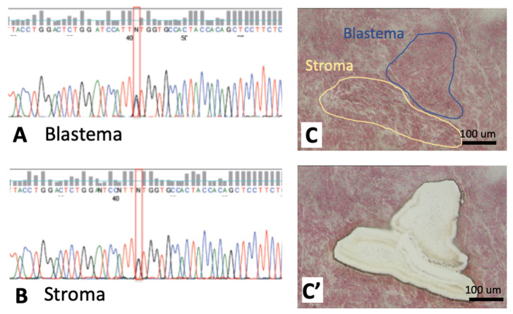
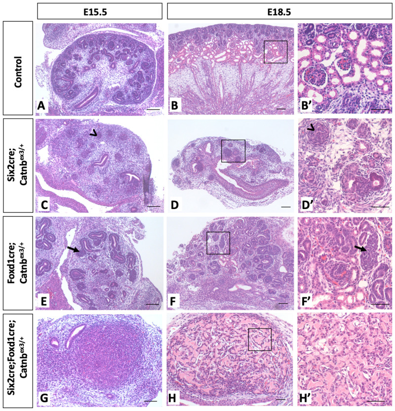
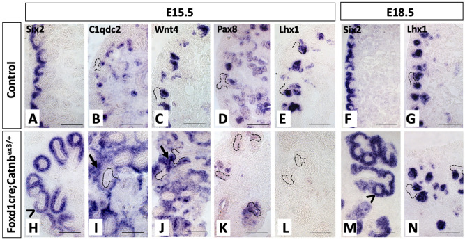
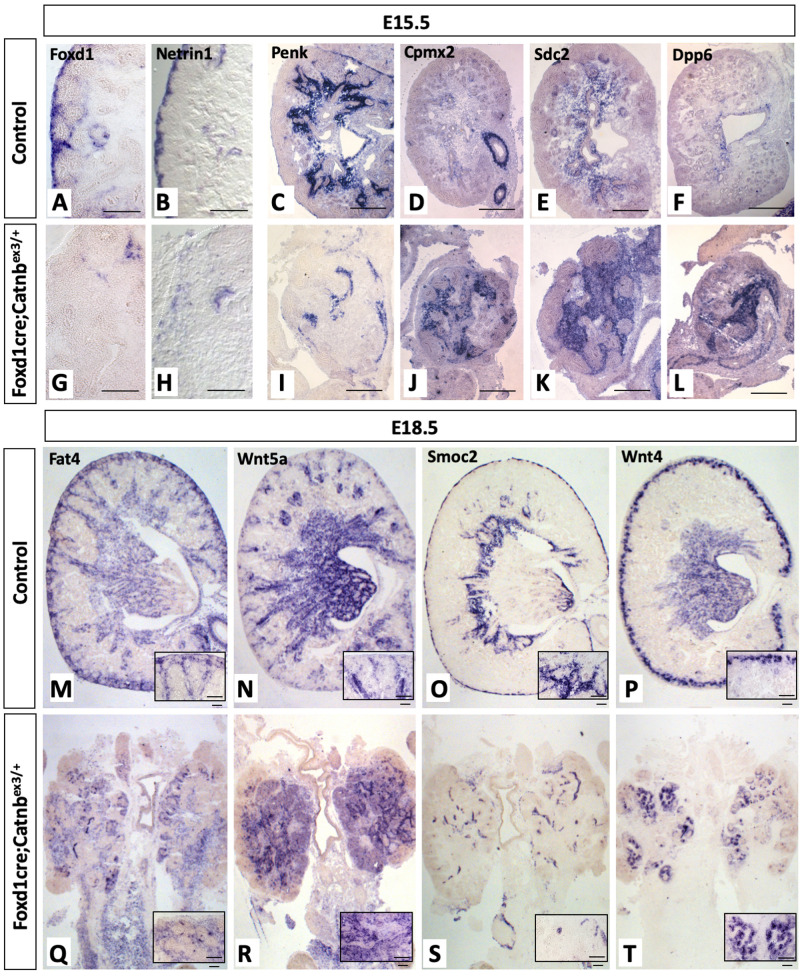
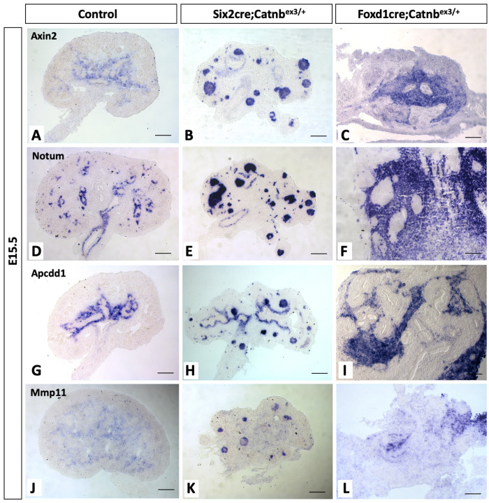
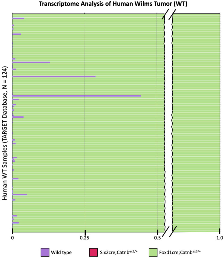
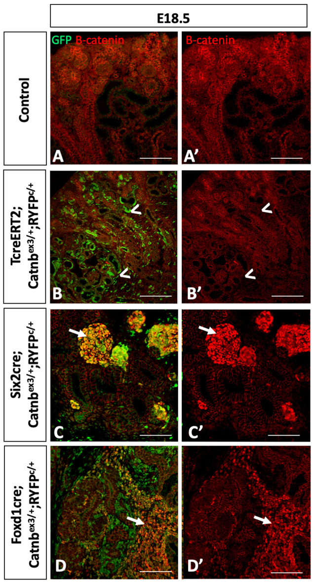
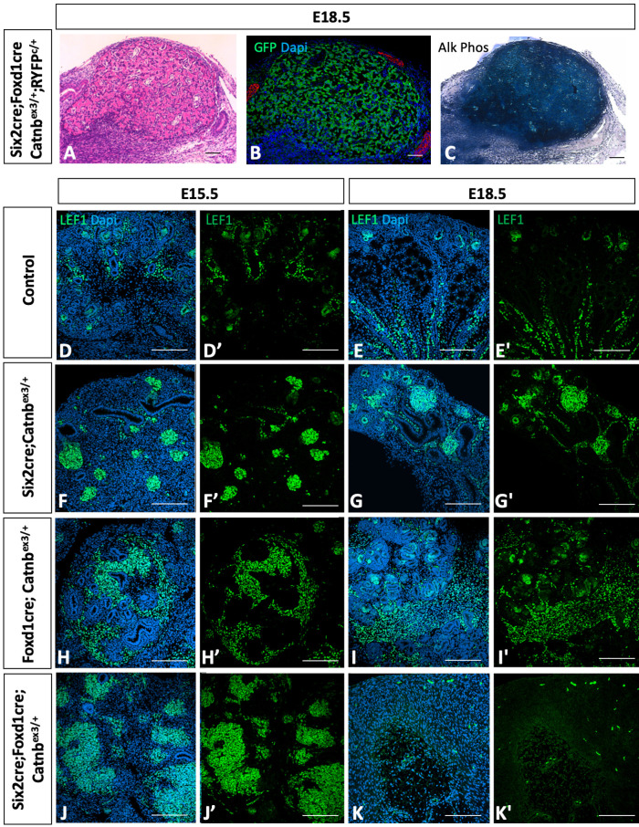
Wilms' tumor (WT) morphologically resembles the embryonic kidney, consisting of blastema, epithelial and stromal components, suggesting tumors arise from the dysregulation of normal development. β-Catenin activation is observed in a significant proportion of WTs; however, much remains to be understood about how it contributes to tumorigenesis. Although activating β-catenin mutations are observed in both blastema and stromal components of WT, current models assume that activation in the blastemal lineage is causal. Paradoxically, studies performed in mice suggest that activation of β-catenin in the nephrogenic lineage results in loss of nephron progenitor cell (NPC) renewal, a phenotype opposite to WT. Here, we show that activation of β-catenin in the stromal lineage non-autonomously prevents the differentiation of NPCs. Comparisons of the transcriptomes of kidneys expressing an activated allele of β-catenin in the stromal or nephron progenitor cells reveals that human WT more closely resembles the stromal-lineage mutants. These findings suggest that stromal β-catenin activation results in histological and molecular features of human WT, providing insights into how alterations in the stromal microenvironment may play an active role in tumorigenesis.

Keywords: Renal development; Renal interstitium; Stroma; Wilms' tumor; β-Catenin.


Conflict of interest statement

Competing interestsThe authors declare no competing or financial interests.

Figures

Fig. 1.
Fig. 1.
Both blastemal and stromal components of human Wilms’ tumor carry CTNNB1-activating mutations. (A-C′) Sequence reads of DNA extracted from blastema (A) and stroma/interstium (B) isolated using a laser microcapture dissection (C,C′) are shown from a representative tumor, demonstrating that both cell populations carry the same CTNNB1 point mutation. Two additional tumors analyzed show the same frame shift in the second tumor and the same in/del in the third (data not shown). Scale bars: 100 μm.
Fig. 2.
Fig. 2.
Activation of β-catenin in different lineages of the developing kidney severely perturbs nephrogenesis, with stromal activation resulting in abnormal maintenance of NPCs and disrupted MET. (A-Hʹ) In comparison with wild-type kidneys (A-B′), Six2cre;Catnbex3/+ kidneys (C-D′) show early loss of NPCs and lack of MET, whereas Foxd1cre;Catnbex3/+ kidneys (E-F') show abnormally maintained NPCs, lacking differentiating structures at E15.5 (E, arrow). By E18.5, some NPCs are induced and undergo nephrogenesis, but regions of abnormally maintained NPCs remain in the developing kidney (F′, arrow). (G-H′) Six2cre;Foxd1cre; Catnbex3/+ kidneys show little resemblance to the developing metanephros and form bone-like tissue later in development. Scale bars: 100 μm. n=3 for each timepoint/genotype.
Fig. 3.
Fig. 3.
β-Catenin activation within the nephron progenitor lineage results in premature loss of the nephron progenitor/blastemal cells, while the opposite phenotype is observed in response to β-catenin activation within the stroma. (A-P) In comparison with control kidneys (A-D), Six2cre;Catnbex3/+ mutant kidneys (E,F) show early loss of Six2-positive NPCs with transient expression of Ncam (E) and Lhx1 (F), consistent with a ‘pre-tubular aggregate (PTA)-like state’, as previously published. (G,H) However, by E15.5, these cells no longer express PTA or renal vesicle markers, including Lhx1, Pax8 and Ncam. (I-L) Foxd1cre;Catnbex3/+ show abnormally maintainted NPCs expressing Six2 and Ncam lacking Lhx1 (K) and Pax8 (L). (M-P) Six2cre;Foxd1cre;Catnbex3/+ mutants initially resemble Six2cre;Catnbex3/+ mutants at E12.5 (M-N), then lose expression of Six2 and Ncam-positive NPCs (O-P). Scale bars: 100 μm. n=3 for each timepoint/genotype.
Fig. 4.
Fig. 4.
β-Catenin activation in stromal lineage results in expanded nephron progenitor cells with delayed MET. (A-L) In comparison with control kidneys (A-G), NPCs of Foxd1cre;Catnbex3/+ mutant kidneys (H-L) show abnormal expansion at E15.5, expressing both markers of both self-renewal (H; Six2, arrowhead) and early commitment to differentiation/MET (I, C1qdc2; J, Wnt4; arrows) but lack expression of other MET markers (K, Pax8; L, Lhx1). (M,N) However, by E18.5, Six2-positive NPCs remained expanded (M, arrowhead) and Lhx1-positive structures are present (N), corresponding to histologically identifiable comma and S-shape-like bodies visualized using Hematoxylin and Eosin staining. Scale bars: 100 μm. n=3 for each timepoint/genotype.
Fig. 5.
Fig. 5.
β-Catenin activation in stromal lineage disrupts normal interstitial patterning. (A-T) Stromal markers in control kidneys (A-F,M-P) were compared with Foxd1cre;Catnbex3/+ mutants (G-L,Q-T), which show early loss of the Foxd1+ stromal progenitor population (G) and nephrogenic interstitial markers netrin 1 (H) and Smoc2 (S). Additionally, medullary stromal markers appear ectopically expressed in the cortex, including Cpmx2 (J), Sdc2 (K), Dpp6 (L) and Wnt5a (R), with a loss of expression of the corticomedullary markers Penk (I) and Smoc2 (S). Scale bars: 100 μm. n=3 for each timepoint/genotype.
Fig. 6.
Fig. 6.
β-Catenin activation in either the NPC or stromal lineages drives the expression of genes normally localized to the developing renal interstitium. (C,F,I,L) Foxd1cre;Catnbex3/+ mutants show upregulation of multiple stromal markers compared with controls (A,D,G,J), as expected given the known role of β-catenin in the development of the medullary interstitium. (B,E,H,K) However, these same target genes are strongly upregulated in Six2cre;Catnbex3/+ cells, somewhat unexpectedly given that these cells originate from a separate lineage where this transcriptional program is not active during normal development. Scale bars: 100 μm; n=3 for each timepoint/genotype.
Fig. 7.
Fig. 7.
Human WT shows molecular characteristics similar to mutant mouse kidneys with activation of β-catenin in stromal lineage. RNA-seq on E12.5 wild-type, Six2cre;Catnbex3/+ and Foxd1cre;Catnbex3/+ mutant kidneys (n=3 for each genotype) were compared with human WT RNA-seq data obtained from the publicly available TARGET database (n=124 samples). Using neural network classification, mapping scores ranging from 0 to 1.0 were generated for each human WT sample measuring similarity of expression of the 2806 identified likely direct targets of β-catenin with that of each of the mouse genotypes, with these results showing expression of these genes in the tumor samples was most similar to the Foxd1cre;Catnbex3/+ mouse model (green bars), with a few tumors showing a small degree of similarity to wild-type kidneys (purple bars), and none of the samples showing any significant degree of similarity to the Six2cre;Catnbex3/+ expression profile.
Fig. 8.
Fig. 8.
β-Catenin activation in early metanephric kidney precursors does not result in nuclear β-catenin despite evidence of recombination. (A,B′) β-Catenin expression in control kidneys (A,A′) was compared with TcreERT2;Catnbex3/+;RosaYFPc/+ mutants given 2 mg per 40 g body weight of tamoxifen at E9.5 (B,B′), which demonstrate recombination by the presence of lineage-traced cells (B, arrowheads); however, these cells unexpectedly lack detectable expression of β-catenin (B′, arrowheads). (C-D′) Conversely, strong nuclear expression is observed in Six2cre and Foxd1cre mutant kidneys (arrows). Scale bars: 100 μm. n=3 for each timepoint/genotype.
Fig. 9.
Fig. 9.
β-Catenin activation in dual NPC and stromal lineages results in the development of bone. (A-C) Six2cre;Foxd1cre;Catnbex3/+ mutant kidneys resemble ‘bone-like’ tissue at E18.5 (Hematoxylin and Eosin staining, A), with reporter expression confirming these cells originated from the targeted cell populations (B), and demonstrate strong expression of the bone marker alkaline phosphatase (C). (D-K′) Although Lef-1, a transcription factor previously shown to interact with β-catenin to promote osteoblast activity (Hoeppner et al., 2011; Li et al., 2018), is upregulated in all mutant lines (F-I′), the ‘bone-like’ phenotype is observed only in the Six2cre;Foxd1cre;Catnbex3/+ mutants (J-K′). Scale bars: 100 μm; n=3 for each timepoint/genotype.

Similar articles

Cited by

References

    1. Boivin F. J. and Bridgewater D. (2018). beta-Catenin in stromal progenitors controls medullary stromal development. Am. J. Physiol. Renal. Physiol. 314, F1177-f1187. 10.1152/ajprenal.00282.2017 - DOI - PubMed
    1. Boivin F. J., Sarin S., Dabas P., Karolak M., Oxburgh L. and Bridgewater D. (2016). Stromal beta-catenin overexpression contributes to the pathogenesis of renal dysplasia. J. Pathol. 239, 174-185. 10.1002/path.4713 - DOI - PubMed
    1. Bremnes R. M., Dønnem T., Al-Saad S., Al-Shibli K., Andersen S., Sirera R., Camps C., Marinez I. and Busund L.-T. (2011). The role of tumor stroma in cancer progression and prognosis: emphasis on carcinoma-associated fibroblasts and non-small cell lung cancer. J. Thorac. Oncol. 6, 209-217. 10.1097/JTO.0b013e3181f8a1bd - DOI - PubMed
    1. Charles A. K., Brown K. W. and Berry P. J. (1998). Microdissecting the genetic events in nephrogenic rests and Wilms' tumor development. Am. J. Pathol. 153, 991-1000. 10.1016/S0002-9440(10)65641-6 - DOI - PMC - PubMed
    1. Clark A. G. and Vignjevic D. M. (2015). Modes of cancer cell invasion and the role of the microenvironment. Curr. Opin. Cell Biol. 36, 13-22. 10.1016/j.ceb.2015.06.004 - DOI - PubMed

Publication types

MeSH terms