. 2020 Jun 30;117(26):14948-14957.
doi: 10.1073/pnas.2003266117. Epub 2020 Jun 15.

RNA-dependent sterol aspartylation in fungi

Affiliations

RNA-dependent sterol aspartylation in fungi

Nathaniel Yakobov et al. Proc Natl Acad Sci U S A. .

Abstract

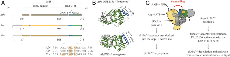
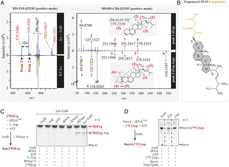
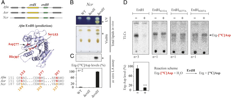
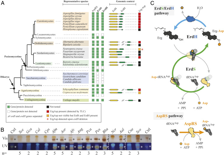
Diverting aminoacyl-transfer RNAs (tRNAs) from protein synthesis is a well-known process used by a wide range of bacteria to aminoacylate membrane constituents. By tRNA-dependently adding amino acids to glycerolipids, bacteria change their cell surface properties, which intensifies antimicrobial drug resistance, pathogenicity, and virulence. No equivalent aminoacylated lipids have been uncovered in any eukaryotic species thus far, suggesting that tRNA-dependent lipid remodeling is a process restricted to prokaryotes. We report here the discovery of ergosteryl-3β-O-l-aspartate (Erg-Asp), a conjugated sterol that is produced by the tRNA-dependent addition of aspartate to the 3β-OH group of ergosterol, the major sterol found in fungal membranes. In fact, Erg-Asp exists in the majority of "higher" fungi, including species of biotechnological interest, and, more importantly, in human pathogens like Aspergillus fumigatus We show that a bifunctional enzyme, ergosteryl-3β-O-l-aspartate synthase (ErdS), is responsible for Erg-Asp synthesis. ErdS corresponds to a unique fusion of an aspartyl-tRNA synthetase-that produces aspartyl-tRNAAsp (Asp-tRNAAsp)-and of a Domain of Unknown Function 2156, which actually transfers aspartate from Asp-tRNAAsp onto ergosterol. We also uncovered that removal of the Asp modifier from Erg-Asp is catalyzed by a second enzyme, ErdH, that is a genuine Erg-Asp hydrolase participating in the turnover of the conjugated sterol in vivo. Phylogenomics highlights that the entire Erg-Asp synthesis/degradation pathway is conserved across "higher" fungi. Given the central roles of sterols and conjugated sterols in fungi, we propose that this tRNA-dependent ergosterol modification and homeostasis system might have broader implications in membrane remodeling, trafficking, antimicrobial resistance, or pathogenicity.

Keywords: DUF2156; aminoacyl-tRNA; ergosterol; fungi; lipid aminoacylation.


Conflict of interest statement

The authors declare no competing interest.

Figures

Fig. 1.
Fig. 1.
Filamentous fungi AspRS-DUF2156, proteins termed ErdSs, are potential lipid aminoacylation factors. (A) In silico analyses predicted Afm, Aor, or Ncr erdS genes to encode proteins composed of an AspRS domain N-terminally fused to a DUF2156 domain. Protein domains were delimited by confronting data obtained from Protein Families database (PFAM) and from multiple alignments as described in SI Appendix, Supplementary Materials and Methods. Critical positively charged residues in the alpha helix (α(+)) separating both GNAT folds are indicated (orange). (B) Comparison of the DUF2156 structure of AlaPGS (alanyl-phosphatidylglycerol synthase, MprF) from P. aeruginosa to the Phyre2 prediction of Afm DUF2156. GNAT I and II subdomains are highlighted in gray and green, respectively, with the positively charged α(+) helix in blue. (C) Schematic representation of the hypothetical ErdS reaction mechanism during which tRNAAsp shift from the AspRS catalytic site (position 1: tRNAAsp aspartylation) to the DUF2156 active site (position 2: Asp transfer from Asp-tRNAAsp onto a lipid substrate). The α(+) helix is indicated in blue, and the active site is indicated in green. Aspartate is represented in orange.
Fig. 2.
Fig. 2.
Identification of an Aspergillus lipid species, whose synthesis requires ErdS. (A) Deletions of erdS from Afm and from Aor were performed by homologous recombination (see SI Appendix for details). The genotypes of the strains are indicated. For Afm, the ΔerdS strain shown corresponds to the strain after excision of the deletion cassette, whereas, for the complemented ΔerdS::PX-erdS strain, the selection marker is still present. For Aor, the ORF of erdS was replaced by a pyrG-containing module and subsequently excised by selection on 5-FOA medium. Complementation was operated by ectopic expression of erdS-ΔDUF, erdS-ΔAspRS, or erdS. (B and C) Total lipids extracted from the different strains described in A were analyzed by TLC and stained with a sulfuric acid/MnCl2 solution. TLC plates were observed either under white light or under UV light. Cultures were done in glucose or xylose containing media; * indicates the LX. (D) Quantification of the TLCs shown in B and C. LX signal (number of pixels) was normalized to that of PE (phosphatidylethanolamine) (LX/PE ratio). All TLCs are representative of at least two independent experiments (n = 2).
Fig. 3.
Fig. 3.
Dissecting ErdS lipid modification mechanism. (A) Schematized modular organization of full-length ErdS (108 kDa, Afm and Aor) and of its variants (Afm) that were expressed in the Sce heterologous model. ErdS-ΔDUF: AspRS standalone domain; ErdS-ΔAspRS: DUF2156 standalone domain (40 kDa); ErdSAAPA: catalytic null of the AspRS moiety (108 kDa). (B) TLC-based analysis of total lipids from Sce expressing ErdS variants described in A and Sce + (ΔD + ΔA) corresponds to the double expression of ErdS-ΔDUF + ErdS-ΔAspRS in Sce. The TLC plate stained with sulfuric acid/MnCl2 was cropped to the area of interest. LX signal (number of pixels) was normalized to that of PE in each case, and the LX/PE ratio is represented in a graph (Right). In the absence of LX, PG (phosphatidylglycerol) becomes visible; thus the PG/LX ratio obtained was considered “background signal” (gray background). However, the brown staining of LX (yellow for PG) made it possible to visually assess the presence of LX even for low LX/PE values. The Student's t test was used to assess the significance of the means of the data; ***P < 0.005. ErdS variants expression was analyzed by Western blot with an anti-Afm DUF2156 polyclonal antibodies (IB:ErdS), and loading control was performed with anti-PGK antibodies (IB:PGK). (C) Schematized reaction of the LA assay, described in Materials and Methods. (D) LX synthesis by the purified recombinant full-length and mutant ErdSs was measured using the LA assay. When mixed, proteins were in equimolar ratios. The [14C]-Asp lipid levels (percent) are provided below each TLC. (E) Verification of tRNA, lipid, and ErdS dependency of LX synthesis using LA assay; +: presence; −: absence. (F) Measurements of lipid X synthesis (ErdS activity) by WT and ΔerdS Aspergillus spp. crude extracts using LA assay; *: LX. The [14C]Asp lipids were revealed using phosphorimaging (DF). TLCs and immunoblots are representative of two to three independent experiments, and the number of replicates (n) is indicated.
Fig. 4.
Fig. 4.
Identification of the ergosteryl-3β-O-l-aspartate produced by ErdS in vivo and in vitro. (A) MS-ESI-QTOF spectrum (positive mode) of a lipid fraction containing LX extracted and purified from an Sce WT strain expressing Afm ErdS (upper spectrum, blue) or not (bottom spectrum, green). Peaks 1 and 2 have been analyzed by MS/MS collision-induced dissociation (CID) QTOF analysis in the positive mode. (B) Chemical structure of ergosteryl-3β-O-l-aspartate (Erg-Ap) corresponding to LX deduced from MS spectra shown in A. (C) The [3H]Erg-Asp synthesis was measured by LA assay in the presence of purified Afm ErdS, pure Sce tRNAAsp, radiolabeled [3H]Erg, and cold Asp in the presence (+) or absence (−) of the enzyme or of the indicated substrates. Tests included addition (+) or not (−) of RNase A. [3H]Erg-Asp is indicated with an asterisk. (D) Erg-[14C]Asp synthesis measured by LA assay using purified Afm ErdS, pure Sce tRNAAsp, radiolabeled [14C]Asp, and indicated sterols. LA assay using total lipids from WT Sce was used as a migration control. The adapted LA assay reactions are displayed (simplified) next to the corresponding TLCs. Radiolabeled compounds are highlighted in red, and the number of independent experiments (n) is indicated.
Fig. 5.
Fig. 5.
Detection and characterization of ErdH in fungi carrying ErdS. (A) Schematic representation of the genomic context of the erdS (yellow) and erdH (for Erg-Asp hydrolase, in green) genes locus from Afm, Aor, and Ncr that highlights that erdS and erdH are found in a divergent orientation. Phyre2-based α/β-hydrolase−like predicted structure of Afm ErdH and active site alignments of Afm, Aor, and Ncr ErdHs. Ser-Asp-His catalytic triads of α/β-hydrolases/lipases (S153, D277, and H307 in Afm ErdH) are displayed and highlighted on the structure prediction and the alignment. (B) Total lipids from WT, ∆erdS, and ∆erdH Ncr strains were separated by TLC and stained with sulfuric acid/MnCl2 and observed under UV or visible light (n = 3); * indicates Erg-Asp. (C) In vitro measurements of Erg-[14C]Asp synthesis by LA assay using protein extracts from the WT, ∆erdS, and ∆erdH Ncr strain protein extracts, using pure Sce tRNAAsp as a substrate (n = 2). (D) In vitro measurement of the Erg-[14C]Asp hydrolase activities of purified recombinant WT (n = 3) or catalytic mutant Ncr ErdHs (n = 1). (C and D) The Student's t test was used to determine significance of the means of the data; ***P < 0.005.
Fig. 6.
Fig. 6.
The Erg-Asp lipid metabolic enzymes ErdS and ErdH are conserved across “higher” fungi. (A) The presence (green square) or absence (white squares) of a regular AspRS, ErdS (yellow), or ErdH (green) genes across fungal species is indicated, and the organization of the locus is shown; // indicates that erdS and erdH genes are interspaced. (B) TLC-based analysis of total lipids extracted from 15 species among the representative ones displayed in A. The TLC plates (number of replicates indicated for each strain) stained with sulfuric acid/MnCl2 solution and observed under white (Vis) or UV light were cropped to the area of interest; * indicates Erg-Asp synthesis (red squares in A) as revealed by the detection of a dark brown band with mobility equivalent to control Erg-Asp. Gca: Geotrichum candidum; Cal: Candida albicans; Cps: Candida parapsilosis; Afv: Aspergillus flavus; Ang: Aspergillus niger; Pca: Penicillium camemberti; Pex: Penicillium expansum; Bba: Beauveria bassiana; Aal: Alternaria alternata; Sco: Schizophyllum commune. (C) A schematic model representing the turnover between Erg-Asp synthesis, and hydrolysis. The green arrow shows ErdS activities (Asp-tRNAAsp production and Asp modification of Erg), and the blue arrow indicates the ErdH-dependent hydrolysis of Erg-Asp. All ErdS-containing fungi always possess a canonical AspRS to ensure Asp-tRNAAsp production for protein synthesis (AspRS pathway).

References

    1. Bohuszewicz O., Liu J., Low H. H., Membrane remodelling in bacteria. J. Struct. Biol. 196, 3–14 (2016). - PMC - PubMed
    1. Fozo E. M., Rucks E. A., The making and taking of lipids: The role of bacterial lipid synthesis and the harnessing of host lipids in bacterial pathogenesis. Adv. Microb. Physiol. 69, 51–155 (2016). - PubMed
    1. Singh A., Del Poeta M., Lipid signalling in pathogenic fungi. Cell. Microbiol. 13, 177–185 (2011). - PMC - PubMed
    1. Fields R. N., Roy H., Deciphering the tRNA-dependent lipid aminoacylation systems in bacteria: Novel components and structural advances. RNA Biol. 15, 480–491 (2018). - PMC - PubMed
    1. Ibba M., Soll D., Aminoacyl-tRNA synthesis. Annu. Rev. Biochem. 69, 617–650 (2000). - PubMed

Publication types

MeSH terms

LinkOut - more resources