Review
. 2020 Jun 15;13(6):dmm044164.
doi: 10.1242/dmm.044164.

Understanding the impact of fibroblast heterogeneity on skin fibrosis

Affiliations
Review

Understanding the impact of fibroblast heterogeneity on skin fibrosis

Michelle F Griffin et al. Dis Model Mech. .

Abstract

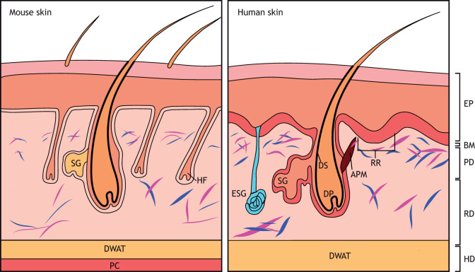
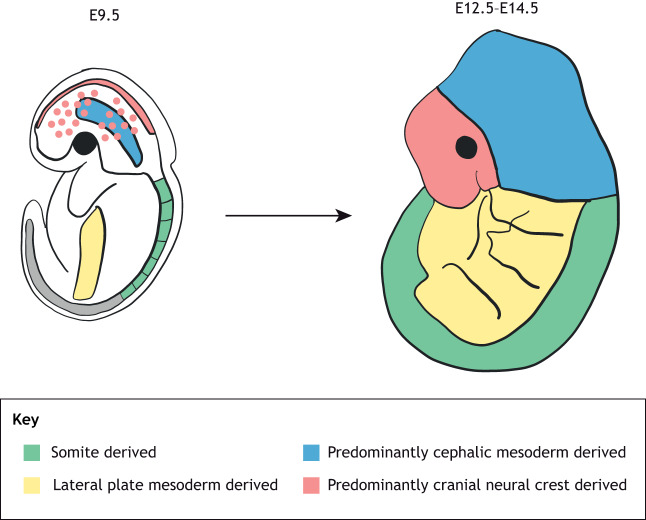
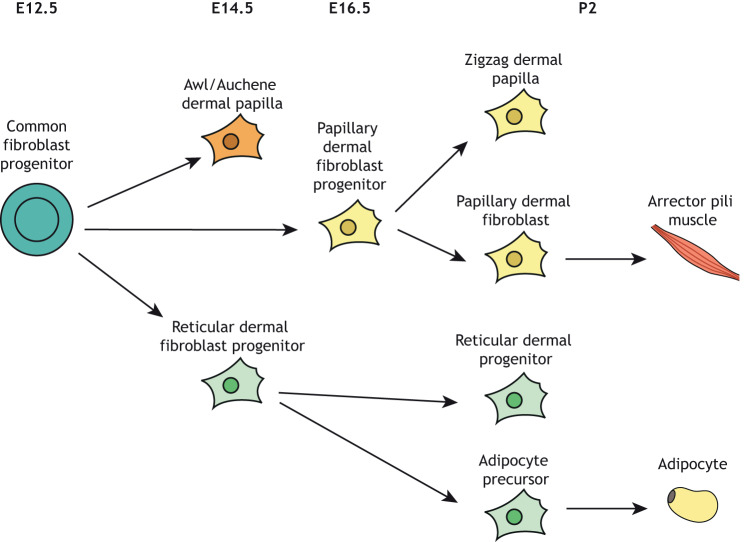
Tissue fibrosis is the deposition of excessive extracellular matrix and can occur as part of the body's natural wound healing process upon injury, or as a consequence of diseases such as systemic sclerosis. Skin fibrosis contributes to significant morbidity due to the prevalence of injuries resulting from trauma and burn. Fibroblasts, the principal cells of the dermis, synthesize extracellular matrix to maintain the skin during homeostasis and also play a pivotal role in all stages of wound healing. Although it was previously believed that fibroblasts are homogeneous and mostly quiescent cells, it has become increasingly recognized that numerous fibroblast subtypes with unique functions and morphologies exist. This Review provides an overview of fibroblast heterogeneity in the mammalian dermis. We explain how fibroblast identity relates to their developmental origin, anatomical site and precise location within the skin tissue architecture in both human and mouse dermis. We discuss current evidence for the varied functionality of fibroblasts within the dermis and the relationships between fibroblast subtypes, and explain the current understanding of how fibroblast subpopulations may be controlled through transcriptional regulatory networks and paracrine communications. We consider how fibroblast heterogeneity can influence wound healing and fibrosis, and how insight into fibroblast heterogeneity could lead to novel therapeutic developments and targets for skin fibrosis. Finally, we contemplate how future studies should be shaped to implement knowledge of fibroblast heterogeneity into clinical practice in order to lessen the burden of skin fibrosis.

Keywords: Dermis; Fibroblast heterogeneity; Scarring; Skin fibrosis; Wound healing.


Conflict of interest statement

Competing interestsThe authors declare no competing or financial interests.

Figures

Fig. 1.
Fig. 1.
Schematic illustration of the skin in mouse and humans. Left: murine skin structure. Mouse skin has a high density of fibroblasts (blue and purple). The panniculus carnosus is under the hypodermis. Right: human skin structure. Human skin structure differs from that of mouse. The epidermis is thicker and forms ingrowths called rete ridges (RR). Hair follicle density in human skin is lower than in mouse. APM, arrector pili muscle; BM, basement membrane; DP, dermal papillae; DS, dermal sheath; DWAT, dermal white adipose tissue; EP, epidermis; ESG, eccrine sweat gland; HD, hypodermis; HF, hair follicle; PC, panniculus carnosus; PD, papillary dermis; RD, reticular dermis; SG, sebaceous gland. Adapted with permission from Rippa et al. (2019). This image is not published under the terms of the CC-BY license of this article. For permission to reuse, please see Rippa et al. (2019).
Fig. 2.
Fig. 2.
Schematic to show the embryonic origin of dermal fibroblasts. The origins of the dermis from different sites of the body are different. The dorsum dermis originates primarily from the somite, the ventrum dermis from the lateral plate, the cranial dermis from the cephalic mesoderm and the face dermis from the neural crest. E, embryonic day. Adapted with permission from Thulabandu et al. (2018). This image is not published under the terms of the CC-BY license of this article. For permission to reuse, please see Thulabandu et al. (2018).
Fig. 3.
Fig. 3.
Schematic of the dermal fibroblast lineages. All fibroblasts originate from a common fibroblast progenitor. In mouse, this process starts at E12.5. Papillary dermal fibroblast progenitors give rise to zig-zag dermal papilla and papillary dermal fibroblasts. Reticular dermal fibroblast progenitors give rise to reticular dermal fibroblasts and adipocytes. E, embryonic day; P, postnatal day. Adapted with permission from Driskell et al. (2013). This image is not published under the terms of the CC-BY license of this article. For permission to reuse, please see Driskell et al. (2013).

References

    1. Akhmetshina A., Palumbo K., Dees C., Bergmann C., Venalis P., Zerr P., Horn A., Kireva T., Beyer C., Zwerina J. et al. (2012). Activation of canonical Wnt signalling is required for TGF-β-mediated fibrosis. Nat. Commun. 3, 735 10.1038/ncomms1734 - DOI - PMC - PubMed
    1. Assassi S., Swindell W. R., Wu M., Tan F. D., Khanna D., Furst D. E., Tashkin D. P., Jahan-Tigh R. R., Mayes M. D., Gudjonsson J. E. et al. (2015). Dissecting the heterogeneity of skin gene expression patterns in systemic sclerosis. Arthritis Rheumatol. 67, 3016-3026. 10.1002/art.39289 - DOI - PMC - PubMed
    1. Bayat A., Mcgrouther D. A. and Ferguson M. W. (2003). Skin scarring. BMJ 326, 88-92. 10.1136/bmj.326.7380.88 - DOI - PMC - PubMed
    1. Bock O., Schmid-Ott G., Malewski P. and Mrowietz U. (2006). Quality of life of patients with keloid and hypertrophic scarring. Arch. Dermatol. Res. 297, 433-438. 10.1007/s00403-006-0651-7 - DOI - PubMed
    1. Cañedo-Dorantes L. and Cañedo-Ayala M. (2019). Skin acute wound healing: a comprehensive review. Int. J. Inflam. 2019, 3706315 10.1155/2019/3706315 - DOI - PMC - PubMed

Publication types

MeSH terms