. 2020 Jun 15;11(1):3034.
doi: 10.1038/s41467-020-16669-9.

Structure and mechanism of DNA delivery of a gene transfer agent

Affiliations

Structure and mechanism of DNA delivery of a gene transfer agent

Pavol Bárdy et al. Nat Commun. .

Abstract

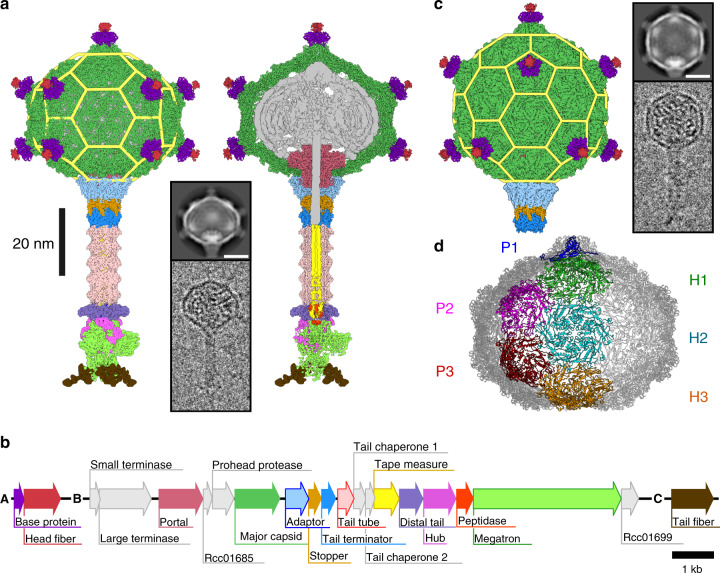
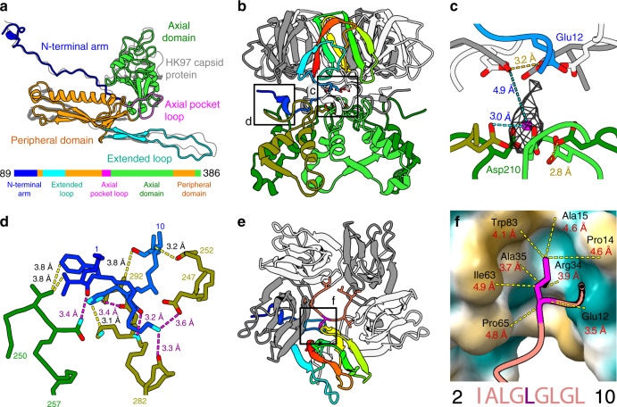
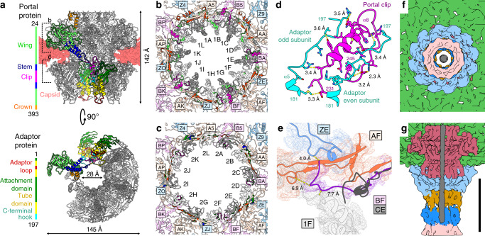
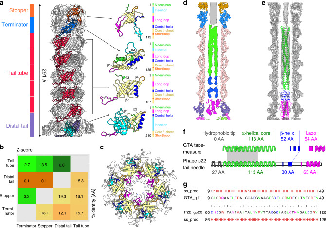
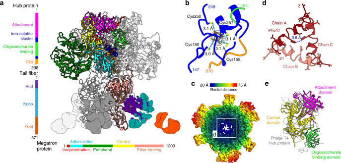
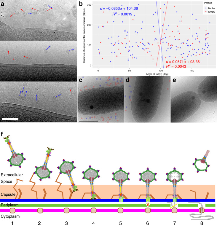
Alphaproteobacteria, which are the most abundant microorganisms of temperate oceans, produce phage-like particles called gene transfer agents (GTAs) that mediate lateral gene exchange. However, the mechanism by which GTAs deliver DNA into cells is unknown. Here we present the structure of the GTA of Rhodobacter capsulatus (RcGTA) and describe the conformational changes required for its DNA ejection. The structure of RcGTA resembles that of a tailed phage, but it has an oblate head shortened in the direction of the tail axis, which limits its packaging capacity to less than 4,500 base pairs of linear double-stranded DNA. The tail channel of RcGTA contains a trimer of proteins that possess features of both tape measure proteins of long-tailed phages from the family Siphoviridae and tail needle proteins of short-tailed phages from the family Podoviridae. The opening of a constriction within the RcGTA baseplate enables the ejection of DNA into bacterial periplasm.


Conflict of interest statement

The authors declare no competing interests.

Figures

Fig. 1
Fig. 1. Structure of the RcGTA particle and organization of segments of the R. capsulatus genome encoding protein components of RcGTA particles.
a Cryo-EM reconstruction of a native particle of RcGTA from R. capsulatus strain DE442 calculated from 42,242 particle images. The left part of the panel shows the complete particle, whereas on the right the front half of the particle has been removed to show DNA and internal proteins. Individual proteins in the density map are colored according to the gene map in panel b. Yellow mesh highlights the structural organization of capsid proteins within the RcGTA head. The inset shows an example of a two-dimensional class average and an electron micrograph of an RcGTA particle. The scale bar within the inset represents 20 nm. b Gene map of three genome segments encoding fourteen structural proteins of RcGTA particles. c Cryo-EM reconstruction of an RcGTA particle from R. capsulatus strain DE442 with T = 3 quasi-icosahedral head. The reconstruction is based on 1076 particle images. The structure is at the scale of those shown in panel a. The inset shows an example of a two-dimensional class average and an electron micrograph of RcGTA particle with an icosahedral head. Scale bar represents 20 nm. d Organization of capsomers in the oblate capsid of RcGTA. Capsomers forming one fifth of the capsid are highlighted in different colors and marked with P for pentamer and H for hexamer.
Fig. 2
Fig. 2. Structure of the major capsid protein and head fibers of RcGTA.
a The major capsid protein of RcGTA has the HK97 fold. The structure of the capsid protein of phage HK97 (PDB 1OHG, shown in gray) is superimposed onto that of RcGTA. The domain organization of the protein is shown in the sequence diagram at the bottom of the panel. b The interaction of base proteins of the head spike with axial domains of major capsid proteins. One subunit of the base protein is rainbow-colored from the N-terminus in blue to the C-terminus in red; the other four subunits are shown in gray and white. The axial domains of the five major capsid proteins are differentiated by shades of green. Details highlighted with black squares are shown in higher magnification in panels c and d. c Coordination of a putative cation by sidechains of Asp210 of major capsid proteins and Glu12 of base proteins strengthens the attachment of base proteins to the RcGTA head. The sidechains of residues interacting with the putative cation are shown in stick representation. Proteins are colored as in b. d Detail of the interaction of the N-terminus of the base protein (blue) with two subunits of major capsid proteins differentiated by being colored in olive and green. Selected interatomic distances are indicated by dashed yellow lines, salt bridges are highlighted with magenta lines. e Top view of a pentamer of base proteins with attached N-terminus of head fiber protein, shown in salmon. The isoleucine and repetition of the leucine residues enable the binding of the head fiber to the pentamer of head spike base proteins. Detail of the indicated interaction is shown in f. f Interaction of leucine 6 of the head fiber, shown in magenta, with the hydrophobic pocket of the head fiber base protein, which is shown as a molecular surface. Yellow indicates a hydrophobic surface and turquoise a charged surface. Distances between selected atoms are indicated. The repetitive sequence from the N-terminus of the head fiber protein is shown at the bottom of the panel.
Fig. 3
Fig. 3. Portal and adaptor complexes.
a Side and bottom views of portal and adaptor complexes with one subunit of the portal protein and two subunits of the adaptor protein highlighted in different colors. One of the adaptor proteins is highlighted with a black outline. Five subunits of portal protein and four subunits of adaptor proteins were removed from the bottom view. The domains of the selected subunits are color-coded as indicated in the sequence diagrams on the left. Differences in the structures of adaptor loops (red) of neighboring adaptor proteins enable reduction of the tail symmetry from twelvefold to six-fold. b, c Interactions of wing domain of portal proteins (b) and attachment domain of adaptor proteins (c) with capsid. Alternating subunits of portal (b) and adaptor (c) complexes are shown in white and gray. Residues of portal and adaptor proteins that interact with the capsid are highlighted in bright colors according to which domain they belong to. The portal and adaptor complexes interact with capsid proteins in three different orientations, which are distinguished by magenta, orange, and blue. Residues of capsid proteins that bind to portal and adaptor proteins are highlighted in bright colors. Subunit are labelled according to PDB 6TBA. d Interactions between the clip domain of the portal protein (magenta) and C-terminal hooks of adaptor proteins (cyan). Sidechains of interacting residues are shown in stick representations. e Unique structure of the N-terminus of capsid protein subunit BF (magenta) interacting with the portal complex (light gray). Capsid protein CE (gray), which does not interact with the portal complex, was superimposed onto the BF subunit. The N-terminus of the CE subunit clashes with the surrounding structures. f, g The end of the double-stranded DNA positioned in the RcGTA neck does not bind to the surrounding proteins of the native RcGTA. f View of tail along its axis towards the center of the head. g View of neck region with front half removed. DNA is shown in gray, tail tube proteins in pink, tail terminator proteins in blue, stopper proteins in dark orange, adaptor proteins in cyan, portal proteins in purple, and capsid proteins in green. Scale bar 10 nm.
Fig. 4
Fig. 4. Structure of the RcGTA tail.
a The tube of the RcGTA tail is formed by the stopper, tail terminator, tail tube, and distal tail proteins. On the right side, the proteins are shown in cartoon representations with β-strands forming the core of the proteins in yellow, N-termini in green, short loops in orange, long loops in magenta, insertion loops in cyan, and central helices in blue. b Structural similarity (upper left) and sequence identity (bottom right) of RcGTA tail proteins. Z-scores were calculated using the DALI server. Values higher than two indicate that the compared proteins are similar. c Superposition of hexamers of stopper proteins of RcGTA, colored as in a, and phage SPP1 (PDB 5A20_EF), in gray. The sidechains of residues that form the bottlenecks in tails of RcGTA and SPP1 are shown in stick representation. The long loop of the stopper protein of RcGTA (magenta) does not reach as close to the center of the channel as that of SPP1. d Central slice through cryo-EM map of RcGTA tail. The parts of the density belonging to RcGTA proteins are color-coded as in panel a. The density in the central channel is color-coded according to the domains of tape measure protein shown in panel f. e Cryo-EM map of RcGTA tail with fitted tail proteins and tail-needle protein of P22 in cartoon representation. The P22 tail-needle model is color-coded according domains shown in panel f. f Tape measure protein of RcGTA is structurally similar to tail-needle protein (PDB 2POH) of phage P22 from the family Podoviridae. Diagrams of secondary structure elements of the two proteins are shown. α-helices are indicated by wiggly lines and β-strands by broad colored lines. The N-terminal region (grey), responsible for the attachment of the needle protein to the tip of P22 tail, is missing from the RcGTA protein. The gray rectangle indicates the position of the sequence displayed in panel g. g Sequence and secondary structure alignment of 41 residues from coiled-coil regions of the RcGTA tape measure protein and phage P22 tail-needle protein computed using HHpred.
Fig. 5
Fig. 5. Structure of the RcGTA baseplate.
a Side-view of the RcGTA baseplate. Domains of one hub and one megatron protein are colored according to the sequence diagrams shown on the left and at the bottom of the panel. The iron–sulfur cluster in the hub protein is shown as dark red spheres. Electron densities of tail fibers are shown. The density of one of the fibers is colored according to domains as indicated in the sequence diagram on the right of the panel. b Detail of iron–sulfur cluster coordinated by four cysteines of hub protein. The electron density map of the cluster is stronger than that corresponding to the surrounding proteins. Distances between sulfur atoms of cysteine sidechains (yellow) and iron ions (red) are indicated. c Cryo-EM map of the RcGTA baseplate viewed along its axis towards the head is rainbow-colored based on the distance from the threefold axis of the structure. The inset shows detail of the constriction of the central channel formed by iris/penetration domains of megatron proteins. d The iris/penetration domains from three subunits of megatron proteins are differentiated by shades of red. Interatomic distances between sidechains of Phe17 are indicated. e Domain swapping among baseplate proteins of RcGTA and phage T4. Attachment and oligosaccharide-binding domains of the RcGTA hub protein and the central domain of the megatron protein of RcGTA can be superimposed onto the central hub protein of bacteriophage T4 shown in gray (PDB 1K28).
Fig. 6
Fig. 6. Mechanism of DNA delivery by RcGTA.
a, b RcGTA particles attach to cells in random orientations. a Cryo-electron micrographs of particles of RcGTA attached to cells of R. capsulatus. The panel includes images from three biological replicates. Orientations of RcGTA tails are highlighted with arrows. Blue indicates genome-containing particles and red empty ones. A cross next to a particle indicates that its tail is not visible in the projection image. Black dots are fiducial markers. Scale bar represents 200 nm. b Distribution of orientations of tails of RcGTA particles (x-axis) versus distance of capsid center from outer cell membrane (y-axis). The tail orientation has values from 0°, when the tail points at the membrane, to 180°, when the tail points away from membrane. The particles are oriented randomly. ce Cells of R. capsulatus are heterogeneous in their capacity to bind RcGTA. Electron micrographs of three R. capsulatus cells from the same experiment using multiplicity of Rif-transferring RcGTA of 0.0002. Some cells were covered with numerous RcGTA particles (c), some had tens of them attached (d), whereas the remaining ones only attracted a few (e). Native and empty RcGTA particles are highlighted with blue and red circles, respectively. f Model of RcGTA-mediated DNA delivery. (1) Free particle. (2) RcGTA attaches to the cell capsule by the head fibers. (3) Particle reorients by the binding of tail fibers to outer membrane receptors. (4) Particle attaches to the membrane by putative receptor-binding domains of the baseplate. (5) Penetration of the outer membrane by iris/penetration domain of megatron protein. (6) Ejection of cell-wall peptidase into periplasm enables degradation of cell wall. (7) Ejection of tape measure protein with DNA to periplasmic space. (8) Uptake of DNA by cell competence system.

References

    1. Lang AS, Westbye AB, Beatty JT. The distribution, evolution, and roles of gene transfer agents in prokaryotic genetic exchange. Annu. Rev. Virol. 2017;4:87–104. - PubMed
    1. Dang H, Li T, Chen M, Huang G. Cross-ocean distribution of Rhodobacterales bacteria as primary surface colonizers in temperate coastal marine waters. Appl. Environ. Microbiol. 2008;74:52–60. - PMC - PubMed
    1. Roux S, et al. Ecogenomics and potential biogeochemical impacts of globally abundant ocean viruses. Nature. 2016;537:689–693. - PubMed
    1. Lang AS, Beatty JT. Genetic analysis of a bacterial genetic exchange element: the gene transfer agent of Rhodobacter capsulatus. Proc. Natl Acad. Sci. USA. 2000;97:859–864. - PMC - PubMed
    1. Fogg PCM. Identification and characterization of a direct activator of a gene transfer agent. Nat. Commun. 2019;10:595. - PMC - PubMed

Publication types

MeSH terms

LinkOut - more resources