. 2020 Aug;30(8):678-692.
doi: 10.1038/s41422-020-0356-z. Epub 2020 Jun 15.

Boceprevir, GC-376, and calpain inhibitors II, XII inhibit SARS-CoV-2 viral replication by targeting the viral main protease

Affiliations

Boceprevir, GC-376, and calpain inhibitors II, XII inhibit SARS-CoV-2 viral replication by targeting the viral main protease

Chunlong Ma et al. Cell Res. 2020 Aug.

Abstract

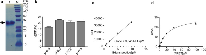
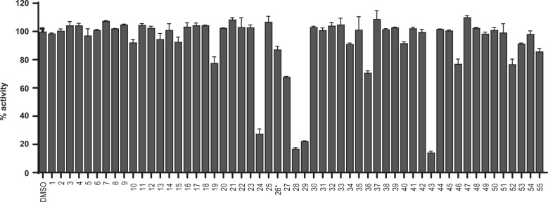
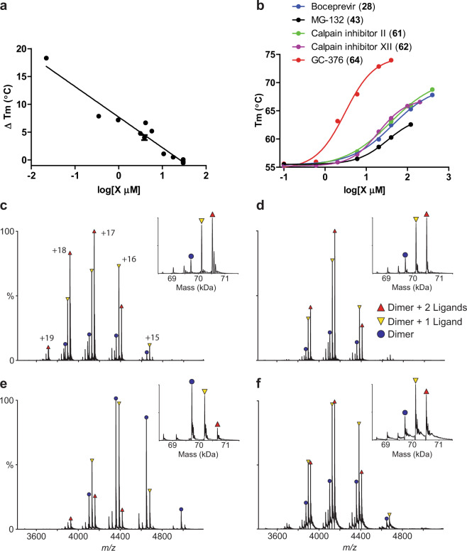
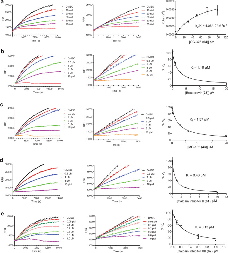
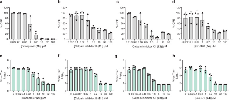
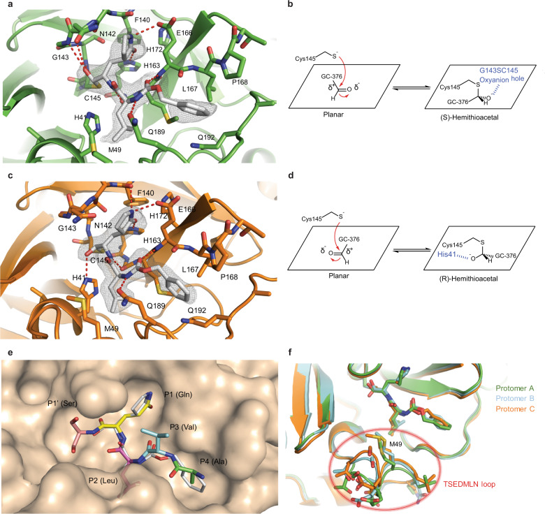
A new coronavirus SARS-CoV-2, also called novel coronavirus 2019 (2019-nCoV), started to circulate among humans around December 2019, and it is now widespread as a global pandemic. The disease caused by SARS-CoV-2 virus is called COVID-19, which is highly contagious and has an overall mortality rate of 6.35% as of May 26, 2020. There is no vaccine or antiviral available for SARS-CoV-2. In this study, we report our discovery of inhibitors targeting the SARS-CoV-2 main protease (Mpro). Using the FRET-based enzymatic assay, several inhibitors including boceprevir, GC-376, and calpain inhibitors II, and XII were identified to have potent activity with single-digit to submicromolar IC50 values in the enzymatic assay. The mechanism of action of the hits was further characterized using enzyme kinetic studies, thermal shift binding assays, and native mass spectrometry. Significantly, four compounds (boceprevir, GC-376, calpain inhibitors II and XII) inhibit SARS-CoV-2 viral replication in cell culture with EC50 values ranging from 0.49 to 3.37 µM. Notably, boceprevir, calpain inhibitors II and XII represent novel chemotypes that are distinct from known substrate-based peptidomimetic Mpro inhibitors. A complex crystal structure of SARS-CoV-2 Mpro with GC-376, determined at 2.15 Å resolution with three protomers per asymmetric unit, revealed two unique binding configurations, shedding light on the molecular interactions and protein conformational flexibility underlying substrate and inhibitor binding by Mpro. Overall, the compounds identified herein provide promising starting points for the further development of SARS-CoV-2 therapeutics.


Conflict of interest statement

J.W. and C.M. are inventors of a pending patent that claims the use of the identified compounds for COVID-19.

Figures

Fig. 1
Fig. 1. SARS-CoV-2 Mpro expression and characterization.
a SDS-PAGE of His-tagged Mpro (lane 1); Lane M, protein ladder; the calculated molecular weight of the His-tagged Mpro is 34,861 Da. b Reaction buffer optimization: 250 nM His-tagged Mpro was diluted into three reaction buffers with different pH values. c An Edans standard curve was generated to convert RFU to the amount of the cleaved substrate (nM). d Michaelis–Menten plot of 200 nM His-tagged Mpro with various concentrations of FRET substrate in pH 6.5 reaction buffer. The best-fit Vmax = 31.7 ± 1.6 nM/s; Km = 28.2 ± 3.4 µM, and the calculated kcat/Km = 5,624 s−1 M−1.
Fig. 2
Fig. 2. Screening of known protease inhibitors against SARS-CoV-2 Mpro using the FRET assay.
20 µM of compounds (26 was tested at 2 µM) was pre-incubated with 100 nM of SARS-CoV-2 Mpro for 30 min at 30 °C, and then 10 µM FRET substrate was added to reaction mixture to initiate the reaction. The reaction was monitored for 2 h. The initial velocity was calculated by linear regression using the data points from the first 15 min of the reaction. The calculated initial velocity with each compound was normalized to DMSO control. The results are average ± standard deviation of two repeats.
Fig. 3
Fig. 3. Binding of inhibitors to SARS-CoV-2 Mpro using thermal shift binding assay and native mass spectrometry.
a Correlation of inhibition efficacy (IC50) with ΔTm from thermal shift binding assay. Data in Table 2 were used for the plot. The r2 of fitting is 0.94. b Dose-dependent melting temperature (Tm) shift. Native MS reveals binding of SARS-CoV-2 Mpro to GC-376 (64) (c), calpain inhibitor II (61) (d), calpain inhibitor XII (62) (e), and boceprevir (28) (f). All ligand concentrations are 12.5 µM except (e), which is 25 µM. Peaks are annotated for dimer (blue circle), dimer with one bound ligand (yellow down triangle), and dimer with two bound ligands (red up triangle). Other minor signals are truncated dimers, which bind ligands at the same ratios. Charge states are annotated in c, and insets show the deconvolved zero-charge mass distribution.
Fig. 4
Fig. 4. Proteolytic reaction progression curves of Mpro in the presence or the absence of compounds.
a–e In the kinetic studies, 5 nM Mpro was added to a solution containing various concentrations of protease inhibitors and 20 µM FRET substrate to initiate the reaction. The reaction was then monitored for 4 h. The left column shows the reaction progression up to 4 h; middle column shows the progression curves for the first 90 min, which were used for curve fitting to generate the plot shown in the right column. Detailed methods were described in “Materials and methods” section. GC-376 (64) (a); boceprevir (28) (b); MG-132 (43) (c); calpain inhibitor II (61) (d); calpain inhibitor XII (62) (e).
Fig. 5
Fig. 5. Antiviral potency of boceprevir (28), calpain inhibitors II (61), XII (62), and GC-376 (64), in CPE and VYR assays.
ad Primary CPE assay results of boceprevir (28), calpain inhibitors II (61), XII (62), and GC-376 (64). eh Secondary VYR assay results of boceprevir (28), calpain inhibitors II (61), XII (62), and GC-376 (64). Each dot represents the average of three repeats from one experiment, and each assay was repeated in three different experiments. The curve fittings were shown in Supplementary information, Fig. S2.
Fig. 6
Fig. 6. Molecular recognition of GC-376 (64) by SARS-CoV-2 Mpro.
Complex of SARS-CoV-2 Mpro and GC-376 (64) with protomer A (a, b) and protomer C (c, d). Unbiased Fo–Fc map, shown in gray, is contoured at 2σ. Hydrogen bonds are shown as red dashed lines. e Surface representation of SARS-CoV-2 Mpro + GC-376 (64) (white) superimposed with the SARS-CoV Mpro natural, N-terminal substrate (PDB ID: 2Q6G, with residues P1′–P4 in different colors). The SARS-CoV-2 Mpro cleaves between the P1′ and P1 residues. f Superimposition of the three protomers in the asymmetric subunit of SARS-CoV-2 Mpro with GC-376 (64). Significant conformational flexibility is observed, particularly in the TSEDMLN loop.

Update of

Comment in

References

    1. Mahase E. Coronavirus: covid-19 has killed more people than SARS and MERS combined, despite lower case fatality rate. BMJ. 2020;368:m641. - PubMed
    1. Lu R, et al. Genomic characterisation and epidemiology of 2019 novel coronavirus: implications for virus origins and receptor binding. Lancet. 2020;395:565–574. - PMC - PubMed
    1. Wu A, et al. Genome composition and divergence of the novel coronavirus (2019-nCoV) originating in China. Cell Host Microbe. 2020;27:325–328. - PMC - PubMed
    1. Coronaviridae Study Group of the International Committee on Taxonomy of Viruses. The species Severe acute respiratory syndrome-related coronavirus: classifying 2019-nCoV and naming it SARS-CoV-2. Nat. Microbiol. 5, 536–544 (2020). - PMC - PubMed
    1. Tang B, et al. An updated estimation of the risk of transmission of the novel coronavirus (2019-nCov) Infect. Dis. Model. 2020;5:248–255. - PMC - PubMed

Publication types

MeSH terms

Substances

LinkOut - more resources