Review
. 2020 Jul;27(7):605-614.
doi: 10.1038/s41594-020-0438-0. Epub 2020 Jun 15.

Targeted protein degradation as a powerful research tool in basic biology and drug target discovery

Affiliations
Review

Targeted protein degradation as a powerful research tool in basic biology and drug target discovery

Tao Wu et al. Nat Struct Mol Biol. 2020 Jul.

Abstract

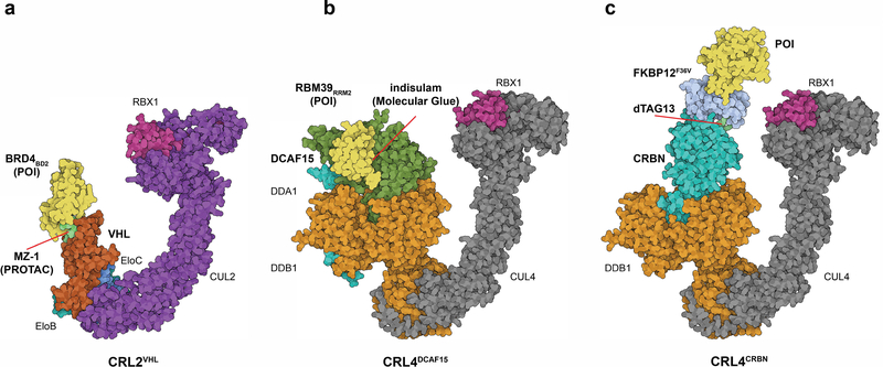
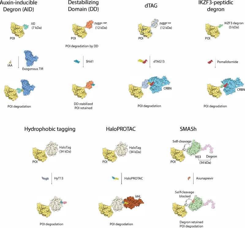
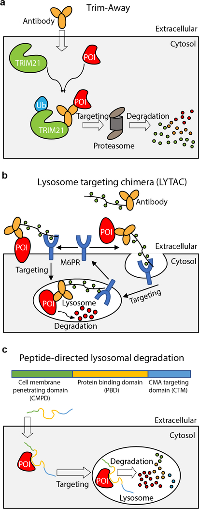
Controlled perturbation of protein activity is essential to study protein function in cells and living organisms. Small molecules that hijack the cellular protein ubiquitination machinery to selectively degrade proteins of interest, so-called degraders, have recently emerged as alternatives to selective chemical inhibitors, both as therapeutic modalities and as powerful research tools. These systems offer unprecedented temporal and spatial control over protein function. Here, we review recent developments in this field, with a particular focus on the use of degraders as research tools to interrogate complex biological problems.


Conflict of interest statement

Competing Interests statement E.S.F. is a founder, scientific advisory board (SAB) member and equity holder of Civetta Therapeutics. E.S.F. is a SAB member and equity holder of C4 Therapeutics. E.S.F. is or has consulted for to Novartis, AbbVie, Astellas, Deerfield, EcoR1 and Pfizer. The Fischer lab receives or has received research funding from Novartis, Deerfield and Astellas.

Figures

Fig. 1.
Fig. 1.
Schematic representations of key concepts in protein degradation. a, Bifunctional degrader (PROTAC). Model of CRL2VHL E3 ligase in complex with MZ-1 (PROTAC) and BRD4BD2 (as a model POI) PDB: 5T35 and 5N4W (chain A and R). b, ‘Molecular glue’ degrader. Model of CRL4DCAF15 E3 ligase in complex with indisulam and RBM39 (POI). PDB: 6Q0R (chain B, C and D), PDB: 4A0C (chain C and D) and PDB: 4A0K (chain C). c, dTAG system as an example for a tagging strategy. Model of CRL4CRBN E3 ligase in complex with dTAG-13 recruiting FKBP12F36V-BRD4BD1 (POI). PDB models as in B, FKBP12 PDB: 1FKJ. Structure visualization created with the Illustrate program
Fig. 2.
Fig. 2.
An overview of degradation tag fusion strategies for targeted protein degradation. Please refer to descriptions in the main text. POI based on BRD4 as an example, PDB: 6BOY; Auxin-inducible Degron based on TIR/IAA structure, PDB: 2P1Q (chain C), TIR structure, PDB: 2P1Q (chain B); Destabilzing Domain FKBP12 structure, PDB: 1FKJ; dTAG CRBN structure, PDB: 6BOY; IKZF3 tag based on structure of IKZF1 ZF2, PDB: 6H0F (chain C); HaloTag based apo HaloTag structure, PDB: 5UY1; VHL structure, PDB: 5T35 (chain D); NS3 protease structure in SMASh, PDB: 4WF8; NS4A degron based structure of NS4A peptide (shorter than the length of degron), PDB: 2OBO (chainB); Structure visualizations created with theIllustrate program.
Fig. 3.
Fig. 3.
Schematic representations of targeted protein degradation strategies mediated by macromolecular conjugates. a, Trim-away. b, Lysosome targeting chimera. c, Peptide-directed lysosomal degradation.
Fig. 4.
Fig. 4.
Validation methods for targeted protein degradation at different stages of the ubiquitin-proteasome pathway. P97 PDB: 5FTJ; model of CRL4CRBN E3 ligase apo structure and structure with BRD4 created with PDB: 4A0C (chain C and D), PDB: 4A0K (chain C) and 6BOY (chain B and C); Human 26S proteasome PDB: 5GJR;Ubiquitin PDB: 1UBQ;POI BRD4 PDB: 6BOY (chain C). Structure visualizations created with the Illustrate program.

References

    1. Ciechanover A Intracellular protein degradation: from a vague idea, through the lysosome and the ubiquitin-proteasome system, and onto human diseases and drug targeting (Nobel lecture). Angew Chem Int Ed Engl 44, 5944–67 (2005). - PubMed
    1. Dikic I Proteasomal and Autophagic Degradation Systems. Annu Rev Biochem 86, 193–224 (2017). - PubMed
    1. Zheng N & Shabek N Ubiquitin Ligases: Structure, Function, and Regulation. Annu Rev Biochem 86, 129–157 (2017). - PubMed
    1. King RW, Glotzer M & Kirschner MW Mutagenic analysis of the destruction signal of mitotic cyclins and structural characterization of ubiquitinated intermediates. Mol Biol Cell 7, 1343–57 (1996). - PMC - PubMed
    1. Takeuchi J, Chen H, Hoyt MA & Coffino P Structural elements of the ubiquitin-independent proteasome degron of ornithine decarboxylase. Biochem J 410, 401–7 (2008). - PubMed

Publication types

MeSH terms