

Mechanisms of innate preconditioning towards ischemia/anoxia tolerance: Lessons from mammalian hibernators
- PMID: 32542230
- PMCID: PMC7295161
Mechanisms of innate preconditioning towards ischemia/anoxia tolerance: Lessons from mammalian hibernators
Abstract
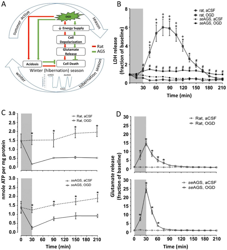
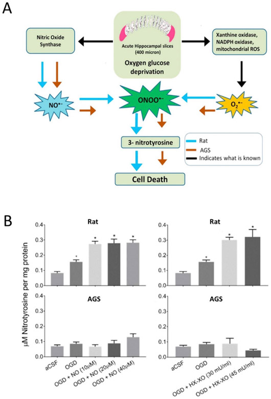
Hibernating mammals exhibit an innate physiological ability to withstand dramatic fluctuations in blood flow that occurs during hibernation and arousal or experimental models of ischemia reperfusion without significant damage. These innate adaptations are of significance particularly to organs that are highly susceptible to energy deprivation, such as the brain and the heart. Among vertebrates, the arctic ground squirrel (AGS) is a species that tolerates ischemic/anoxic insult. During the process of entering hibernation, a state of prolonged torpor, the AGS undergoes a profound decrease in respiratory rate, heart rate, blood flow, cerebral perfusion, and body temperature (Tb). The reduced level of blood flow during torpor resembles an ischemic state, albeit without energy deficit. During the process of arousal or emergence from torpor, however, when Tb, respiratory rate, heart rate, and blood flow rapidly returns to pre-torpid levels, the rapid return of cerebral blood flow mimics aspects of reperfusion such as is seen after stroke or cardiac arrest. This sublethal ischemic/reperfusion insult experienced by AGS during the process of arousal may precondition AGS to tolerate otherwise lethal ischemic/reperfusion injury induced in the laboratory. In this review, we will summarize some of the mechanisms implemented by mammalian hibernators to combat ischemia/anoxia tolerance.
Conflict of interest statement
Conflict of interest statement The authors declare that they have no conflicts of interest.
Figures
References
-
- Anderson TR, Jarvis CR, Biedermann AJ, Molnar C, Andrew RD (2005) Blocking the anoxic depolarization protects without functional compromise following simulated stroke in cortical brain slices. J Neurophysiol 93:963–979. - PubMed
-
- Astrup J, Symon L, Branston NM, Lassen NA (1977) Cortical evoked potential and extracellular K+ and H+ at critical levels of brain ischemia. Stroke 8:51–57. - PubMed
-
- Barnes BM (1989) Freeze avoidance in a mammal: body temperatures below 0 degree C in an Arctic hibemator. Science 244:1593–1595. - PubMed
Grants and funding
LinkOut - more resources
Full Text Sources