Review
. 2020 Jun 26;134(12):1333-1356.
doi: 10.1042/CS20200279.

Microvascular disease in chronic kidney disease: the base of the iceberg in cardiovascular comorbidity

Affiliations
Review

Microvascular disease in chronic kidney disease: the base of the iceberg in cardiovascular comorbidity

Uwe Querfeld et al. Clin Sci (Lond). .

Abstract

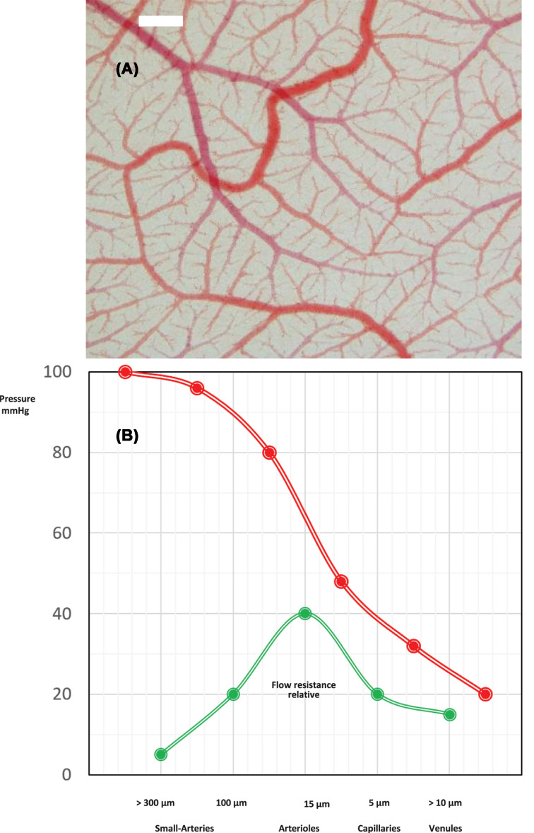
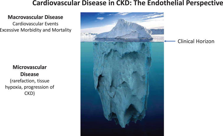
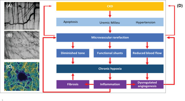
Chronic kidney disease (CKD) is a relentlessly progressive disease with a very high mortality mainly due to cardiovascular complications. Endothelial dysfunction is well documented in CKD and permanent loss of endothelial homeostasis leads to progressive organ damage. Most of the vast endothelial surface area is part of the microcirculation, but most research in CKD-related cardiovascular disease (CVD) has been devoted to macrovascular complications. We have reviewed all publications evaluating structure and function of the microcirculation in humans with CKD and animals with experimental CKD. Microvascular rarefaction, defined as a loss of perfused microvessels resulting in a significant decrease in microvascular density, is a quintessential finding in these studies. The median microvascular density was reduced by 29% in skeletal muscle and 24% in the heart in animal models of CKD and by 32% in human biopsy, autopsy and imaging studies. CKD induces rarefaction due to the loss of coherent vessel systems distal to the level of smaller arterioles, generating a typical heterogeneous pattern with avascular patches, resulting in a dysfunctional endothelium with diminished perfusion, shunting and tissue hypoxia. Endothelial cell apoptosis, hypertension, multiple metabolic, endocrine and immune disturbances of the uremic milieu and specifically, a dysregulated angiogenesis, all contribute to the multifactorial pathogenesis. By setting the stage for the development of tissue fibrosis and end organ failure, microvascular rarefaction is a principal pathogenic factor in the development of severe organ dysfunction in CKD patients, especially CVD, cerebrovascular dysfunction, muscular atrophy, cachexia, and progression of kidney disease. Treatment strategies for microvascular disease are urgently needed.

Keywords: Microcirculation; capillary; cardiovascular disease; chronic kidney disease; endothelial dysfunction; hypertension.


Conflict of interest statement

The authors declare that there are no competing interests associated with the manuscript.

Figures

Figure 1
Figure 1. The microcirculatiory network
(A) Vascular network of the chorioallantoic membrane. In-vivo microphotography, bar = 100 μm. (B) Functional anatomy: pressure and relative flow resistance for coronary vessels of different sizes. The main flow resistance and pressure decrease is located in the arteriolar section of the coronary tree (modified from [190]).
Figure 2
Figure 2. The ‘iceberg delusion’ may prevent appreciation of the magnitude of CVD in CKD patients
Most of the vast endothelial surface area is part of the microcirculation. Considering the entire vasculature, CKD affects first and foremost the microcirculation in a silent, subclinical process. When macrovascular involvement and CVD events appear on the clinical horizon, these late events can be viewed as the tip of the iceberg of CKD-induced vascular disease.
Figure 3
Figure 3. Microvascular disease in CKD
(A,B,C) Microvascular rarefaction in-vivo. In-vivo microscopic view (40×) of the microvasculature of an area (350 × 250 μm) of the murine M. cremaster. Panel (A) shows microvessels with a normal density of a control mouse, panel (B) the avascular area of a mouse with severe adenine-induced uremia. (C) Optical coherence tomography angiography, vessel density map of the superior vascular plexus of the retina in a patient with CKD stage 5. Avascular zone of the fovea shown in black, patchy distribution of areas of capillary rarefaction shown in blue (taken from Yeung et al. [75]). (D) A simplified scheme of the pathogenesis of microvascular disease in CKD.

References

    1. Bonetti P.O., Lerman L.O. and Lerman A. (2003) Endothelial dysfunction: a marker of atherosclerotic risk. Arterioscler. Thromb. Vasc. Biol. 23, 168–175 10.1161/01.ATV.0000051384.43104.FC - DOI - PubMed
    1. Fliser D. et al. . (2011) The dysfunctional endothelium in CKD and in cardiovascular disease: mapping the origin(s) of cardiovascular problems in CKD and of kidney disease in cardiovascular conditions for a research agenda. Kidney Int. Suppl. 1, 6–9 10.1038/kisup.2011.6 - DOI - PMC - PubMed
    1. Levy B.I., Ambrosio G., Pries A.R. and Struijker-Boudier H.A. (2001) Microcirculation in hypertension: a new target for treatment? Circulation 104, 735–740 10.1161/hc3101.091158 - DOI - PubMed
    1. Virdis A. et al. . (2014) Evaluation of microvascular structure in humans: a ‘state-of-the-art’ document of the Working Group on Macrovascular and Microvascular Alterations of the Italian Society of Arterial Hypertension. J. Hypertens. 32, 2120–2129, 10.1097/HJH.0000000000000322 - DOI - PubMed
    1. Pries A.R., Secomb T.W. and Gaehtgens P. (2000) The endothelial surface layer. Pflug. Arch. 440, 653–666 10.1007/s004240000307 - DOI - PubMed

MeSH terms