

Comparison of regional flortaucipir PET with quantitative tau immunohistochemistry in three subjects with Alzheimer's disease pathology: a clinicopathological study
- PMID: 32542468
- PMCID: PMC7295920
- DOI: 10.1186/s13550-020-00653-x
Comparison of regional flortaucipir PET with quantitative tau immunohistochemistry in three subjects with Alzheimer's disease pathology: a clinicopathological study
Abstract
Background: The objective of this study was to make a quantitative comparison of flortaucipir PET retention with pathological tau and β-amyloid across a range of brain regions at autopsy.
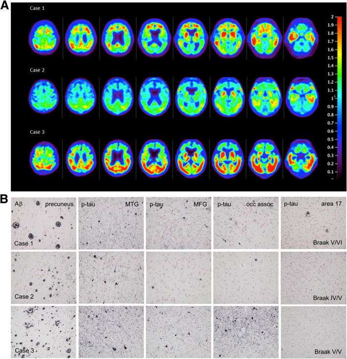
Methods: Patients with dementia (two with clinical diagnosis of AD, one undetermined), nearing the end of life, underwent 20-min PET, beginning 80 min after an injection of ~370 mBq flortaucipir [18F]. Neocortical, basal ganglia, and limbic tissue samples were obtained bilaterally from 19 regions at autopsy and subject-specific PET regions of interest corresponding to the 19 sampled target tissue regions in each hemisphere were hand drawn on the PET images. SUVr values were calculated for each region using a cerebellar reference region. Abnormally phosphorylated tau (Ptau) and amyloid-β (Aβ) tissue concentrations were measured for each tissue region with an antibody capture assay (Histelide) using AT8 and H31L21 antibodies respectively.
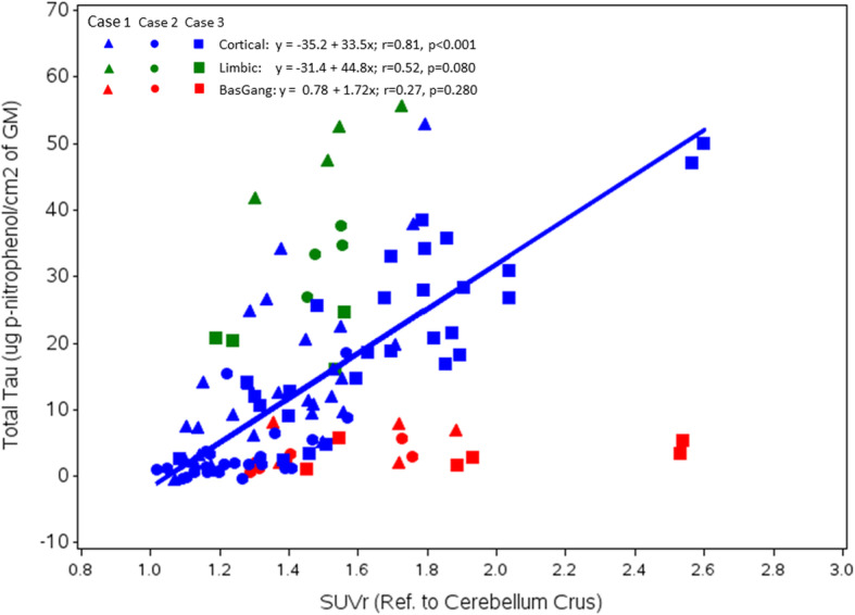
Results: The imaging-to-autopsy interval ranged from 4-29 days. All three subjects had intermediate to high levels of AD neuropathologic change at autopsy. Mean cortical SUVr averaged across all three subjects correlated significantly with the Ptau immunoassay (Pearson r = 0.81; p < 0.0001). When Ptau and Aβ1-42 were both included in the model, the Ptau correlation with flortaucipir SUVr was preserved but there was no correlation of Aβ1-42 with flortaucipir. There was also a modest correlation between limbic (hippocampal/entorhinal and amygdala) flortaucipir SUVr and Ptau (Pearson r = 0.52; p < 0.080). There was no significant correlation between SUVr and Ptau in basal ganglia.
Conclusions: The results of this pilot study support a quantitative relationship between cortical flortaucipir SUVr values and quantitative measures of Ptau at autopsy. Additional research including more cases is needed to confirm the generalizability of these results. Trial registration, NIH Clinicaltrials.gov NCT # 02516046. Registered August 27, 2015. https://clinicaltrials.gov/ct2/show/NCT02516046?term=02516046&draw=2&rank=1.
Keywords: Alzheimer’s; Autopsy; Flortaucipir; Histelide; NFT; PET; PHF; Tau; [18F]-AV-1451.
Conflict of interest statement
MJP, AKA, MDD, MN, IK, ML, PA, MF, ASF, and MAM are full-time employees of Avid Radiopharmaceuticals, a wholly owned subsidiary of Eli Lilly and Company, and are minor stockholders of Eli Lilly and Company. ADJ and AS were employees of Avid at the time this work was performed but are now at Medpace Holdings and University of Pennsylvania, respectively. TGB, TJM, CDK, GES, LIS, AJI, SER, AW, LH, and NC contributed to this work as part of a research contract with Avid.
Figures
References
-
- Nelson PT, Jicha GA, Schmitt FA, Liu H, Davis DG, Mendiondo MS, et al. Clinicopathologic correlations in a large Alzheimer disease center autopsy cohort: neuritic plaques and neurofibrillary tangles "do count" when staging disease severity. J Neuropathol Exp Neurol. 2007;66:1136–1146. doi: 10.1097/nen.0b013e31815c5efb. - DOI - PMC - PubMed
Associated data
Grants and funding
LinkOut - more resources
Full Text Sources
Medical
