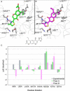
X-Ray Crystallography and Free Energy Calculations Reveal the Binding Mechanism of A2A Adenosine Receptor Antagonists
- PMID: 32542862
- PMCID: PMC7540567
- DOI: 10.1002/anie.202003788
X-Ray Crystallography and Free Energy Calculations Reveal the Binding Mechanism of A2A Adenosine Receptor Antagonists
Abstract
We present a robust protocol based on iterations of free energy perturbation (FEP) calculations, chemical synthesis, biophysical mapping and X-ray crystallography to reveal the binding mode of an antagonist series to the A2A adenosine receptor (AR). Eight A2A AR binding site mutations from biophysical mapping experiments were initially analyzed with sidechain FEP simulations, performed on alternate binding modes. The results distinctively supported one binding mode, which was subsequently used to design new chromone derivatives. Their affinities for the A2A AR were experimentally determined and investigated through a cycle of ligand-FEP calculations, validating the binding orientation of the different chemical substituents proposed. Subsequent X-ray crystallography of the A2A AR with a low and a high affinity chromone derivative confirmed the predicted binding orientation. The new molecules and structures here reported were driven by free energy calculations, and provide new insights on antagonist binding to the A2A AR, an emerging target in immuno-oncology.
Keywords: G protein-coupled receptor (GPCR); adenosine receptors; biophysical mapping (BPM); free energy perturbation (FEP).
© 2020 The Authors. Published by Wiley-VCH Verlag GmbH & Co. KGaA.
Conflict of interest statement
The authors declare no conflict of interest.
Figures


References
-
- Cournia Z., Allen B., Sherman W., J. Chem. Inf. Model. 2017, 57, 2911–2937. - PubMed
-
- Rao S. N., Singh U. C., Bash P. A., Kollman P. A., Nature 1987, 328, 551–554. - PubMed
-
- Jespers W., Isaksen G. V., Andberg T. A. H., Vasile S., Van Veen A., Åqvist J., Brandsdal B. O., Gutiérrez-De-Terán H., J. Chem. Theory Comput. 2019, 15, 5461–5473. - PubMed
-
- Steinbrecher T. B., Dahlgren M., Cappel D., Lin T., Wang L., Krilov G., Abel R., Friesner R., Sherman W., J. Chem. Inf. Model. 2015, 55, 2411–2420. - PubMed
Publication types
MeSH terms
Substances
LinkOut - more resources
Full Text Sources
Research Materials

