Review
. 2020 Jun;10(6):200097.
doi: 10.1098/rsob.200097. Epub 2020 Jun 17.

Bacterial chromosome segregation by the ParABS system

Affiliations
Review

Bacterial chromosome segregation by the ParABS system

Adam S B Jalal et al. Open Biol. 2020 Jun.

Abstract

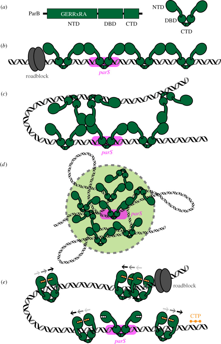
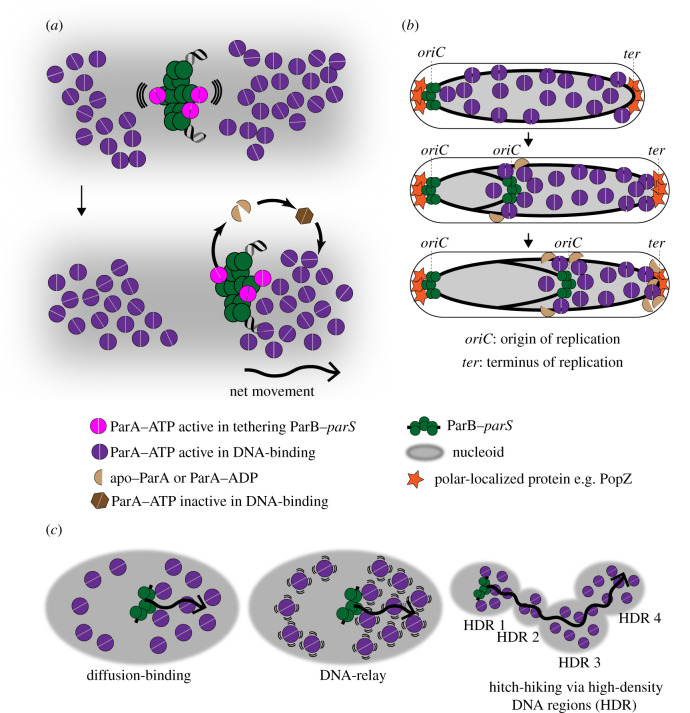
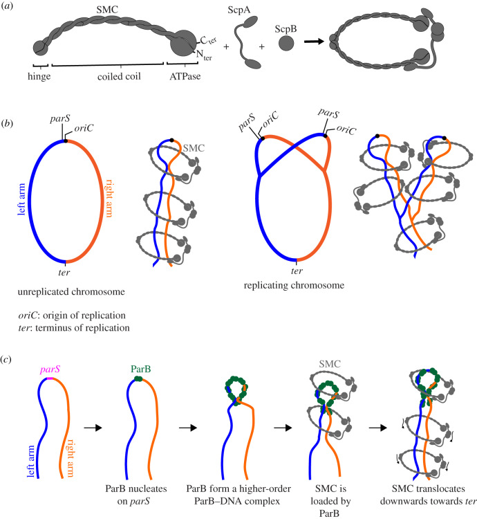
Proper chromosome segregation during cell division is essential in all domains of life. In the majority of bacterial species, faithful chromosome segregation is mediated by the tripartite ParABS system, consisting of an ATPase protein ParA, a CTPase and DNA-binding protein ParB, and a centromere-like parS site. The parS site is most often located near the origin of replication and is segregated first after chromosome replication. ParB nucleates on parS before binding to adjacent non-specific DNA to form a multimeric nucleoprotein complex. ParA interacts with ParB to drive the higher-order ParB-DNA complex, and hence the replicating chromosomes, to each daughter cell. Here, we review the various models for the formation of the ParABS complex and describe its role in segregating the origin-proximal region of the chromosome. Additionally, we discuss outstanding questions and challenges in understanding bacterial chromosome segregation.

Keywords: ParA–ParB–parS; SMC; chromosome maintenance; chromosome organization; chromosome segregation; spreading.


Conflict of interest statement

We declare we have no competing interests.

Figures

Figure 1.
Figure 1.
The assembly of a higher-order ParB–DNA nucleoprotein complex. (a) Chromosomal ParB proteins share a common domain architecture, consisting of an N-terminal domain (NTD), a central DNA-binding domain (DBD) and a C-terminal domain (CTD). The NTD harbours a conserved arginine-rich motif (GERRxRA) that mediates ParB–ParB and ParB–cytidine triphosphate (CTP) interactions. (b) Model 1: ParB spreading by a one-dimensional filamentation. (c) Model 2: ParB spreading by bridging and condensing DNA. (d) Model 3: ParB spreading by caging DNA. (e) Model 4: ParB spreading by sliding on DNA. ParB switches from an open to a closed clamp upon binding to CTP (orange). ParB and parS are coloured green and magenta, respectively. The arrows above the ParB–CTP complexes (e) indicate their progressive sliding on DNA. A tight DNA-binding protein (grey) can unidirectionally block the one-dimensional filamentation or the sliding of ParB on DNA.
Figure 2.
Figure 2.
ParA drives the movement of ParB-bound DNA to segregate plasmids and chromosomes. (a) A diffusion-ratchet model for ParA-mediated transport of ParB-bound DNA. A ParB–DNA complex (green) interacts with ParA–ATP (violet) to tether to the nucleoid (grey), and to stimulate the ATPase activity of ParA. ParA–ATP dimers (violet) bind the nucleoid non-specifically. After ATP hydrolysis, monomers of apo–ParA/ParA–ADP (light brown) no longer bind DNA, thus creating a zone of depletion of ParA–ATP surrounding the ParB–DNA complex. By thermal fluctuation (wavy lines), the ParB–DNA complex moves to the edge of the zone of depletion to rebind ParA–ATP. The initial movement of the ParB–DNA complex in one chosen direction enforces the continued movement in the same direction, resulting in a long-range directional movement of the DNA (see b). The released apo–ParA/ParA–ADP (light brown) rebinds ATP but cannot immediately bind DNA (the dark brown hexagon) until a transition occurs in the ParA–ATP structure. (b) The segregation of the origin-proximal region of the chromosome by the ParABS system. For example, in C. crescentus, one ParB–DNA complex remains at the pole after chromosome replication, while the other moves along the gradient of ParA–ATP, via the diffusion-ratchet mechanism, to the opposite cell pole. The polarly localized proteins (e.g. PopZ, orange) contribute to maintaining the ParA–ATP gradient by sequestering apo–ParA/ParA–ADP away from the nucleoid and to regenerate them at the pole. (c) Other variations of the diffusion-ratchet model have been proposed to include an element of DNA elasticity (i.e. the DNA-relay model) or high-density DNA regions (HDR) (i.e. the hitch-hiking model). A wavy arrow indicates the directional movement of the partition complex.
Figure 3.
Figure 3.
The ParB–DNA nucleoprotein complex recruits SMC to coordinate chromosome segregation and chromosome organization. (a) Components of the bacterial SMC complex. (b) SMC (grey) tethers the two arms (blue and orange) of a circular chromosome together. An SMC–ScpA–ScpB complex can either hold both the left and the right arm of the chromosome within its lumen or two SMC complexes, each encircles one chromosome arm, can handcuff to tether both chromosome arms together. For simplicity, only SMCs entrapping both chromosome arms are shown. SMC probably packages sister chromosomes into individual entities and away from each other, thus minimizing DNA entanglement between replicating chromosomes. (c) A schematic model of how SMC is loaded at parS by ParB and translocates on the chromosome towards the replication terminus (ter). For simplicity, alternative conformations of SMC (ring or rod) are not illustrated; the SMC complex is shown as a generic ring that entraps DNA. Schematic pictures are not drawn to scale.

References

    1. Abeles AL, Friedman SA, Austin SJ. 1985. Partition of unit-copy miniplasmids to daughter cells. III. The DNA sequence and functional organization of the P1 partition region. J. Mol. Biol. 185, 261–272. (10.1016/0022-2836(85)90402-4) - DOI - PubMed
    1. Austin S, Abeles A. 1983. Partition of unit-copy miniplasmids to daughter cells. II. The partition region of miniplasmid P1 encodes an essential protein and a centromere-like site at which it acts. J. Mol. Biol. 169, 373–387. (10.1016/s0022-2836(83)80056-4) - DOI - PubMed
    1. Austin S, Abeles A. 1983. Partition of unit-copy miniplasmids to daughter cells. I. P1 and F miniplasmids contain discrete, interchangeable sequences sufficient to promote equipartition. J. Mol. Biol. 169, 353–372. (10.1016/s0022-2836(83)80055-2) - DOI - PubMed
    1. Mori H, Kondo A, Ohshima A, Ogura T, Hiraga S. 1986. Structure and function of the F plasmid genes essential for partitioning. J. Mol. Biol. 192, 1–15. (10.1016/0022-2836(86)90459-6) - DOI - PubMed
    1. Ireton K, Gunther NW, Grossman AD. 1994. spo0 J is required for normal chromosome segregation as well as the initiation of sporulation in Bacillus subtilis. J. Bacteriol. 176, 5320–5329. (10.1128/jb.176.17.5320-5329.1994) - DOI - PMC - PubMed

Publication types

MeSH terms

Substances

LinkOut - more resources