. 2020 Sep 1;130(9):4798-4810.
doi: 10.1172/JCI132987.

3D model of harlequin ichthyosis reveals inflammatory therapeutic targets

Affiliations

3D model of harlequin ichthyosis reveals inflammatory therapeutic targets

Florence Enjalbert et al. J Clin Invest. .

Abstract

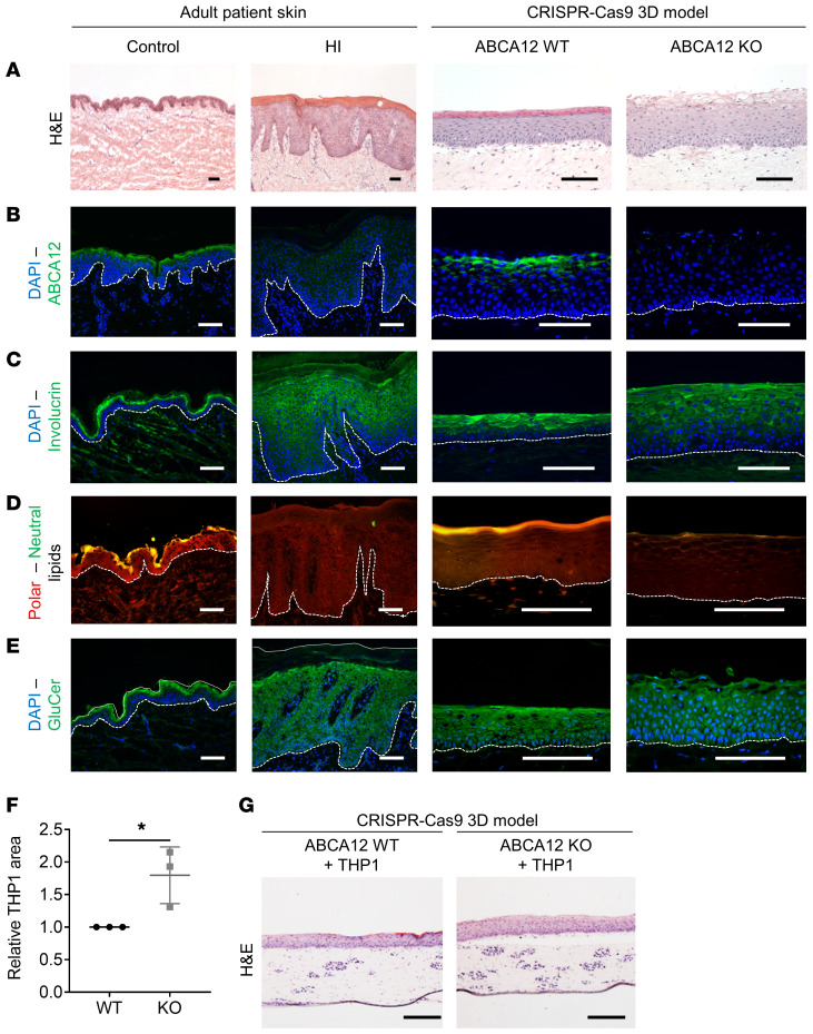
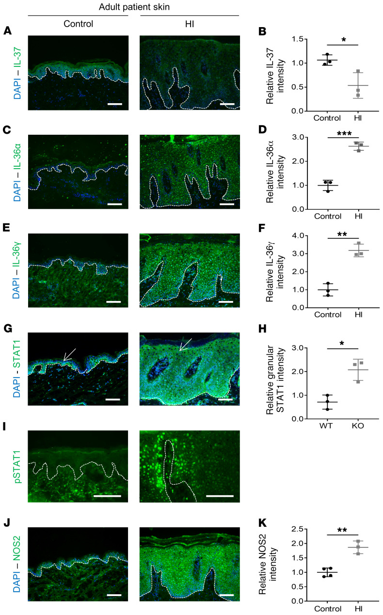
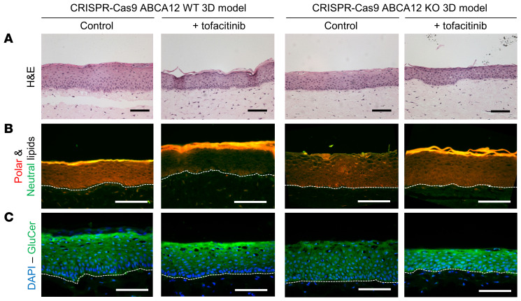
The biology of harlequin ichthyosis (HI), a devastating skin disorder caused by loss-of-function mutations in the gene ABCA12, is poorly understood, and to date, no satisfactory treatment has been developed. We sought to investigate pathomechanisms of HI that could lead to the identification of new treatments for improving patients' quality of life. In this study, RNA-Seq and functional assays were performed to define the effects of loss of ABCA12 using HI patient skin samples and an engineered CRISPR/Cas9 ABCA12 KO cell line. The HI living skin equivalent (3D model) recapitulated the HI skin phenotype. The cytokines IL-36α and IL-36γ were upregulated in HI skin, whereas the innate immune inhibitor IL-37 was strongly downregulated. We also identified STAT1 and its downstream target inducible nitric oxide synthase (NOS2) as being upregulated in the in vitro HI 3D model and HI patient skin samples. Inhibition of NOS2 using the inhibitor 1400W or the JAK inhibitor tofacitinib dramatically improved the in vitro HI phenotype by restoring the lipid barrier in the HI 3D model. Our study has identified dysregulated pathways in HI skin that are feasible therapeutic targets.

Keywords: Dermatology; Genetic diseases; Genetics; Nitric oxide; Skin.


Conflict of interest statement

Conflict of interest: EAO has received research funding from Kamari Pharma in the current fiscal year.

Figures

Figure 1
Figure 1. ABCA12 KO–induced changes in lipid distribution, cellular morphology and growth, and increased inflammatory response in 2D culture.
(A) Representative immunoblot of ABCA12 and GAPDH proteins in ABCA12 WT and KO cell lysates. (B) Representative Nile red–staining images of polar/neutral (red/green channel) lipids in ABCA12 WT and KO cells. Scale bars: 50 μm. (C) Associated quantitative lipid droplet number analysis. Each dot represents the mean of 3 technical replicates. n = 3. Data are represented as mean ± SD. *P ≤ 0.05, unpaired t test. (D) Representative fluorescence staining images of CellMask and DAPI in ABCA12 WT and KO cells. Scale bars: 100 μm. (E) Associated quantitative cell area analysis. Each dot represents the mean of 3 technical replicates. n = 3. Data are represented as mean ± SD. *P ≤ 0.05, unpaired t test. (F) Cell proliferation analysis of ABCA12 WT and KO cells. n = 3. Data are represented as mean ± SD. ****P ≤ 0.0001, 2-way ANOVA with Šidák’s multiple comparisons test. Measurement of secreted (G) IL-1α and (H) CXCL1 in ABCA12 WT and KO supernatants. Each dot represents the mean of 3 technical replicates. n = 3. Mean ± SD. *P ≤ 0.05; **P < 0.01, unpaired t test.
Figure 2
Figure 2. HI skin ABCA12 KO 3D model showed alterations in keratinocyte differentiation, lipid expression pattern, and inflammation.
Representative (A) H&E (bright-field channel) staining images and (B) ABCA12 (green channel), (C) involucrin (green channel), (D) polar/neutral (red/green channel) lipid, (E) GluCer (green channel), and DAPI (blue channel) immunofluorescence staining images of control skin, HI patient skin, and in vitro WT and HI 3D models. Scale bars: 100 μm. (F) Quantitative analysis of relative THP-1 cellular area in the dermis-like layers of ABCA12 WT and KO cells. Each dot represents the mean of relative THP-1 area from 3 independent images. n = 3. Data are represented as mean ± SD. *P ≤ 0.05, unpaired t test. (G) Associated H&E-stained (bright-field channel) representative images. Scale bars: 200 μm.
Figure 3
Figure 3. Transcriptomic profile of HI skin and ABCA12 KO model using RNA-Seq.
Volcano plot of differentially expressed genes between (A) 4 HI skin and 5 normal skin controls and (B) CRISPR/Cas9 ABCA12 KO and WT 3D models. Each red/black dot represents a significantly differentially up- or downregulated gene. (C) GO terms enrichment in differentially expressed genes (upregulated in red, downregulated in black) in HI skin compared with normal skin. (D) Functional protein association network; line thickness indicates the strength of data support. (E) GO terms enrichment in differentially expressed genes (upregulated in red, downregulated in black) in CRISPR/Cas9 ABCA12 KO 3D models compared with control.
Figure 4
Figure 4. Decrease in antiinflammatory response and activation of NOS2 pathway in the in vitro ABCA12 KO 3D model.
(A) qPCR analysis of IL37 in ABCA12 WT and KO 3D models. n = 3. Data are represented as mean ± SD. NS, P = 0.068, unpaired t test. qPCR analysis of (B) SOCS1 and (C) SOCS3 in ABCA12 WT and KO 2D model cell lysates. Each dot represents the mean of 3 technical replicates. n = 3. Data are represented as mean ± SD. **P ≤ 0.01, unpaired t test. (D) Representative immunoblot of p-STAT1 (Y701), total STAT1, and GAPDH proteins in untreated (–) or stimulated (+) with IFN-γ ABCA12 WT and KO cell lysates and (E) associated p-STAT1 quantitative analysis. n = 4. Data are represented as mean ± SD. *P ≤ 0.05, unpaired t test. The p-STAT1 blot was run in parallel, contemporaneously, with total STAT1 and GAPDH blots. (F) Representative NOS2 (green channel) and DAPI (blue channel) staining images of in vitro WT and HI 3D models, and (G) associated quantitative NOS2 analysis. Each dot represents the mean of relative NOS2 intensity from 3 independent images. Scale bars: 100 μm. n = 3. Data are represented as mean ± SD. *P ≤ 0.05, unpaired t test.
Figure 5
Figure 5. Inflammation and activation of the STAT1/NOS2 pathway in HI skin.
Representative (A) IL-37, (C) IL-36α, (E) IL-36γ, (G) STAT1, (I) p-STAT1, (J) and NOS2 (green channel) and DAPI (blue channel) staining images of control skin and HI patient skin. Arrows indicate granular layer. Associated quantitative analysis of (B) IL-37, (D) IL-36α, (F) IL-36γ, (H) granular layer STAT1, and (K) NOS2 protein expression in control skin and HI patient skin. Each dot represents the mean of relative protein intensity from 3 independent images. n = 3 or 4. Data are represented as mean ± SD. *P ≤ 0.05; **P ≤ 0.01; ***P ≤ 0.001, unpaired t test. Scale bars: 100 μm.
Figure 6
Figure 6. NO release caused epidermal acanthosis and inhibition of NOS2 resulted in normalization of the skin barrier in the HI 3D model.
(A) Representative H&E (bright-field channel) images of in vitro WT and HI 3D models untreated (UT) or treated with SNAP compound. (B) Quantitative analysis of intracellular NO in in vitro WT and HI 3D models, with or without 1400 W inhibitor. Each dot represents the mean of 3 technical replicates. n = 3. Data are represented as mean ± SD. **P ≤ 0.01; ***P ≤ 0.001, 2-way ANOVA with Tukey’s multiple comparisons test. Representative (C) H&E (bright-field channel), (D) Lucifer yellow (LY) (green channel), (E) polar/neutral (red/green channel), (F) GluCer (green channel), and DAPI (blue channel) staining images of in vitro WT and HI 3D models from 3 independent biological replicates. Scale bars: 100 μm.
Figure 7
Figure 7. Tofacitinib treatment improved skin-barrier formation in the WT and HI 3D model.
Representative (A) H&E (bright-field channel), (B) polar/neutral (red/green channel), (C) GluCer (green channel), and DAPI (blue channel) staining images of in vitro WT and HI 3D models with or without tofacitinib from 3 independent biological replicates. Scale bars: 100 μm.

Similar articles

Cited by

References

    1. Lee SH, et al. 16-kDa prolactin down-regulates inducible nitric oxide synthase expression through inhibition of the signal transducer and activator of transcription 1/IFN regulatory factor-1 pathway. Cancer Res. 2005;65(17):7984–7992. doi: 10.1158/0008-5472.CAN-05-0631. - DOI - PubMed
    1. Oji V, et al. Revised nomenclature and classification of inherited ichthyoses: results of the First Ichthyosis Consensus Conference in Sorèze 2009. J Am Acad Dermatol. 2010;63(4):607–641. - PubMed
    1. Oji V, Traupe H. Ichthyosis: clinical manifestations and practical treatment options. Am J Clin Dermatol. 2009;10(6):351–364. doi: 10.2165/11311070-000000000-00000. - DOI - PubMed
    1. Kelsell DP, et al. Mutations in ABCA12 underlie the severe congenital skin disease harlequin ichthyosis. Am J Hum Genet. 2005;76(5):794–803. doi: 10.1086/429844. - DOI - PMC - PubMed
    1. Akiyama M, et al. Mutations in lipid transporter ABCA12 in harlequin ichthyosis and functional recovery by corrective gene transfer. J Clin Invest. 2005;115(7):1777–1784. doi: 10.1172/JCI24834. - DOI - PMC - PubMed

Publication types

MeSH terms