. 2020 Jul 27;48(13):7265-7278.
doi: 10.1093/nar/gkaa524.

Disease-associated DNA2 nuclease-helicase protects cells from lethal chromosome under-replication

Affiliations

Disease-associated DNA2 nuclease-helicase protects cells from lethal chromosome under-replication

Benoît Falquet et al. Nucleic Acids Res. .

Abstract

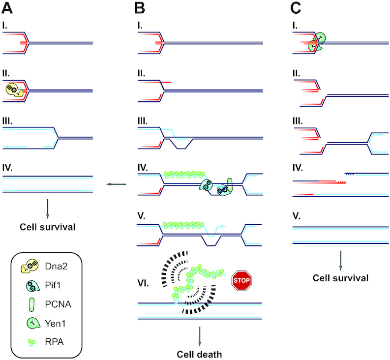
DNA2 is an essential nuclease-helicase implicated in DNA repair, lagging-strand DNA synthesis, and the recovery of stalled DNA replication forks (RFs). In Saccharomyces cerevisiae, dna2Δ inviability is reversed by deletion of the conserved helicase PIF1 and/or DNA damage checkpoint-mediator RAD9. It has been suggested that Pif1 drives the formation of long 5'-flaps during Okazaki fragment maturation, and that the essential function of Dna2 is to remove these intermediates. In the absence of Dna2, 5'-flaps are thought to accumulate on the lagging strand, resulting in DNA damage-checkpoint arrest and cell death. In line with Dna2's role in RF recovery, we find that the loss of Dna2 results in severe chromosome under-replication downstream of endogenous and exogenous RF-stalling. Importantly, unfaithful chromosome replication in Dna2-mutant cells is exacerbated by Pif1, which triggers the DNA damage checkpoint along a pathway involving Pif1's ability to promote homologous recombination-coupled replication. We propose that Dna2 fulfils its essential function by promoting RF recovery, facilitating replication completion while suppressing excessive RF restart by recombination-dependent replication (RDR) and checkpoint activation. The critical nature of Dna2's role in controlling the fate of stalled RFs provides a framework to rationalize the involvement of DNA2 in Seckel syndrome and cancer.


Figures

Figure 1.
Figure 1.
Dna2 is required for the completion of chromosome replication. (A) Representative PFGE experiments with the indicated strains, DNA stained by ethidium bromide. G1-synchronized cells were released into S phase in the presence of nocodazole to arrest cells prior to mitosis. At the indicated experimental stages, genomic DNA was analyzed for fully replicated (gel-resolved) chromosomes by PFGE. Gel-resolved DNA is labelled with chromosome numbers. (B) Southern blot analysis of the gel shown in panel A, probing for chromosome XII. (C) Quantification of Southern blots as shown in panel B to determine the fraction of gel-resolved chromosome XII. Data represent mean values ± SEM (n = 3 independent experiments). (D) Cell-cycle progression analysis by flow cytometry of cells collected as in panel A.
Figure 2.
Figure 2.
Transient replication arrest induces under-replication in the absence of Dna2. (A) Representative PFGE of the indicated strains treated with HU, DNA stained by ethidium bromide. G1-synchronized cells were released into medium containing 200 mM HU for 2 h, followed by drug wash-out and incubation in HU-free medium containing nocodazole to prevent mitosis. At the indicated stages, genomic DNA was analyzed for fully replicated (gel-resolved) chromosomes by PFGE. Gel-resolved DNA is labelled with chromosome numbers. (B) Cell-cycle progression analysis by flow cytometry of cells collected for the experiment shown in panel A. (C) Southern blot analysis of a gel obtained as in panel A, probing for chromosome XII. (D) Quantification of Southern blots as shown in panel C to determine the fraction of gel-resolved chromosome XII. Data represent mean values ± SEM (n = 3 independent experiments). (E) Southern blot analysis of a gel obtained as in panel A, probing for chromosome XIII. (F) Quantification of Southern blots as shown in panel E to determine the fraction of gel-resolved chromosome XIII. Data represent mean values ± SEM (n = 3 independent experiments).
Figure 3.
Figure 3.
Transient RF arrest in the absence of Dna2 is cell-lethal. (A) Cell-cycle progression analysis by flow cytometry of the indicated strains synchronized in G1, treated with 200 mM HU for 2 h and released for 4 h into a drug-free medium. (B) Representative images of dna2Δ pif1-m2 cells treated as in panel A showing single-nucleated cells (I), a double-nucleated cell in G2 (II), a G2 cell with a nucleus at the bud neck (III), and an anaphase cell with an elongated nucleus spanning the bud neck (IV). Scale bar, 5 μm. (C) Quantification of cells (n ≥ 110 cells per strain) observed as in panel B. (D) Cell viability of the indicated strains, assessed by plating efficiency (PE) after synchronization in G1, removal of α-factor, and treatment or not with 200 mM HU for 2 h. Data expressed relative to pif1-m2 cells as mean values ± SD (n = 3 independent experiments).
Figure 4.
Figure 4.
The resolution of under-replicated chromosomes by Yen1 is indispensable in the absence of Dna2. (A) Requirement for YEN1 and MUS81 in dna2Δ pif1-m2 cells. Cells contained a Yen1-expressing plasmid (pYEN1). Lack of growth on 5-FOA indicates an inviable genotype. (B) SIM image of an anaphasic pif1-m2 control cell expressing Yen1-EGFP (green). Scale bar, 5μm. (C) Representative images of anaphasic pif1-m2 (i-ii) and dna2Δ pif1-m2 (iii-vi) cells expressing Yen1-EGFP. Images v and vi show Yen1 foci located on a loop-like structure indicative of rDNA (arrowheads). Scale bar, 5 μm. (D) Quantification of Yen1-EGFP foci per anaphase cell in the indicated strains. Unperturbed samples are from exponentially growing cultures. For HU samples, cells were synchronized in G1, treated with 200 mM HU for 2 h and released into drug-free medium. All data represented as violin plots with the mean ± SD (n = 3 independent experiments, ≥ 99 cells observed per strain and condition). Statistical analysis by one-way analysis of variance (Anova) and post-hoc Tukey multiple comparison test (n.s., non-significant; **P< 0.01; ***P< 0.001). (E) Quantification as in panel D, focused on Yen1 foci between segregating masses of DNA. (F) Representative images of anaphasic dna2Δ pif1-m2 cells with Yen1-EGFP (green) and Nop1-dsRED (magenta) foci on the separating DNA masses (top panel, arrowheads) and within the anaphase tube connecting them (bottom panel). Cells were synchronized in G1, treated with 200 mM HU for 2 h and released into drug-free medium. Scale bar, 5 μm. (G) Quantification of Yen1 and Nop1 co-localization, determined as in panel F. Data are represented as mean values ± SD (n = 3 independent experiments, ≥61 cells scored per strain and condition).
Figure 5.
Figure 5.
Incomplete replication upon Dna2 dysfunction gives rise to Pif1-mediated DNA damage checkpoint activation. (A) Drop assays with the indicated strains on drug-free and HU-containing plates to assess growth and RS-sensitivity. (B) Mitotic time-course experiments with transient RS-treatment of the indicated strains. Cells, synchronized in G1, were released into medium containing 50 mM HU for 2 h, followed by drug wash-out and incubation in drug-free medium with α-factor to prevent entry into a second S phase. Checkpoint activation was monitored by Western blot analysis of Rad53 hyperphosphorylation (black arrowheads). The progression of DNA replication was monitored by flow cytometry (1 and 2 N DNA content are indicated).
Figure 6.
Figure 6.
Pif1-toxicity in Dna2-defective cells maps to homologous recombination-coupled DNA replication. (A) Schematic representation of Pif1 showing the bi-partite helicase domain (grey), and a TLSSAES phosphorylation motif and PCNA-interacting peptide (PIP box) required for homologous recombination-coupled DNA replication. Mutations introduced into these domains are denoted below, see text for details. HD, helicase-dead. (B) Drop assays of serial dilutions on drug-free and HU-containing medium of the indicated strains expressing wild-type Pif1 (+pPIF1), a helicase-dead version of Pif1 (+pPIF1-HD), or containing the empty vector. (C) Drop assays with the indicated strains on drug-free and HU-containing medium assessing growth and sensitivity to RS in the presence or absence of the pif1-R3E and pif1-4a alleles at the endogenous PIF1 locus in multiple independently created strains. (D) dna2-HD pif1-4a cells were assessed for unscheduled G2/M DNA damage-checkpoint activation following transient exposure to RS as in Figure 5, panel B. (E) The mutational analysis of Pif1 strongly implicates homologous recombination-coupled DNA replication at stalled RFs as the pathway generating toxicity by unscheduled DNA damage-checkpoint activation in Dna2-mutant cells.
Figure 7.
Figure 7.
Model of Dna2 fulfilling an essential role at stalled RFs by promoting RF recovery and suppressing inappropriate restart by RDR. (A) Dna2 counteracts RF reversal at stalled RFs (I) by degrading nascent ssDNA or unwinding and degrading the regressed dsDNA arm (II) (6,31). This enables direct resumption of RF progression (III) and promotes the completion of chromosome duplication (IV). (B) Stalled RFs left unprocessed by Dna2 (I) are subject to alternative restart by RDR (II), leading to the formation of a D-loop (III). Pif1 promotes homologous recombination-coupled DNA replication (19,20) by binding PCNA and stimulating DNA synthesis in the context of a D-loop (57) (IV). RDR can facilitate replication completion and cell survival (arrow pointing left). However, migrating D-loops are characterized by frequent nascent strand dissociation (V) (66–69). D-loop collapse and passage of a conventional RF may cause nascent RDR strands to become permanently exposed as ssDNA, resulting in checkpoint-activating structures with long ssDNA branches that conform to Pif1-dependent intermediates recently observed in Dna2-depleted cells (VI) (32). This explains why Dna2 is essential: In the absence of Dna2, inappropriate RDR results in toxic levels of RPA-covered ssDNA, causing DNA damage-checkpoint activation and terminal cell-cycle arrest. (C) In the absence of Dna2 and Pif1, replication remains incomplete, but because stalled RFs are unable to undergo excessive RDR, the DNA damage checkpoint remains silent and cells enter mitosis. This activates Yen1 (I), enabling the resolution of chromosome entanglements at persistent replication intermediates (II). The DNA repair steps downstream of Yen1-cleavage remain to be determined. It is conceivable that arrival of a converging RF (III) facilitates replication of the unbroken sister chromatid and DSB repair on the sister chromatid cleaved by Yen1 (IV), thereby mediating replication completion (V).

Similar articles

Cited by

References

    1. Bae S.H., Choi E., Lee K.H., Park J.S., Lee S.H., Seo Y.S.. Dna2 of Saccharomyces cerevisiae possesses a single-stranded DNA-specific endonuclease activity that is able to act on double-stranded DNA in the presence of ATP. J. Biol. Chem. 1998; 273:26880–26890. - PubMed
    1. Budd M.E., Campbell J.L.. A yeast gene required for DNA replication encodes a protein with homology to DNA helicases. Proc. Natl. Acad. Sci. U.S.A. 1995; 92:7642–7646. - PMC - PubMed
    1. Pinto C., Kasaciunaite K., Seidel R., Cejka P.. Human DNA2 possesses a cryptic DNA unwinding activity that functionally integrates with BLM or WRN helicases. Elife. 2016; 5:e18574. - PMC - PubMed
    1. Lin W., Sampathi S., Dai H., Liu C., Zhou M., Hu J., Huang Q., Campbell J., Shin-Ya K., Zheng L. et al. .. Mammalian DNA2 helicase/nuclease cleaves G-quadruplex DNA and is required for telomere integrity. EMBO J. 2013; 32:1425–1439. - PMC - PubMed
    1. Hoa N.N., Kobayashi J., Omura M., Hirakawa M., Yang S.-H., Komatsu K., Paull T.T., Takeda S., Sasanuma H.. BRCA1 and CtIP Are Both Required to Recruit Dna2 at Double-Strand Breaks in Homologous Recombination. PLoS ONE. 2015; 10:e0124495. - PMC - PubMed

Publication types