Review
. 2020 Sep:96:102-113.
doi: 10.1016/j.reprotox.2020.06.007. Epub 2020 Jun 13.

Retinoids and developmental neurotoxicity: Utilizing toxicogenomics to enhance adverse outcome pathways and testing strategies

Affiliations
Review

Retinoids and developmental neurotoxicity: Utilizing toxicogenomics to enhance adverse outcome pathways and testing strategies

Hao Chen et al. Reprod Toxicol. 2020 Sep.

Abstract

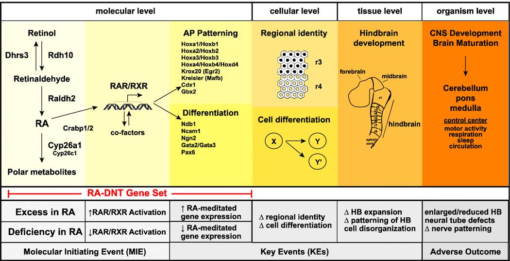
The use of genomic approaches in toxicological studies has greatly increased our ability to define the molecular profiles of environmental chemicals associated with developmental neurotoxicity (DNT). Integration of these approaches with adverse outcome pathways (AOPs), a framework that translates environmental exposures to adverse developmental phenotypes, can potentially inform DNT testing strategies. Here, using retinoic acid (RA) as a case example, we demonstrate that the integration of toxicogenomic profiles into the AOP framework can be used to establish a paradigm for chemical testing. RA is a critical regulatory signaling molecule involved in multiple aspects of mammalian central nervous system (CNS) development, including hindbrain formation/patterning and neuronal differentiation, and imbalances in RA signaling pathways are linked with DNT. While the mechanisms remain unresolved, environmental chemicals can cause DNT by disrupting the RA signaling pathway. First, we reviewed literature evidence of RA and other retinoid exposures and DNT to define a provisional AOP related to imbalances in RA embryonic bioavailability and hindbrain development. Next, by integrating toxicogenomic datasets, we defined a relevant transcriptomic signature associated with RA-induced developmental neurotoxicity (RA-DNT) in human and rodent models that was tested against zebrafish model data, demonstrating potential for integration into an AOP framework. Finally, we demonstrated how these approaches may be systematically utilized to identify chemical hazards by testing the RA-DNT signature against azoles, a proposed class of compounds that alters RA-signaling. The provisional AOP from this study can be expanded in the future to better define DNT biomarkers relevant to RA signaling and toxicity.

Keywords: Alternative model; Azoles; Embryonic stem cells; Human; In vitro; Neurogenesis; Neurotoxicity; Retinoic acid; Retinoids; Transcriptome; Whole embryo culture; Zebrafish.


Figures

Figure 1.
Figure 1.. Retinoic acid metabolism.
The availability of RA is tightly controlled in the mammalian CNS. Mammals are unable to synthesize RA de novo and require intake of vitamin A or other precursors (β-carotenes) from food sources. Vitamin A is converted to retinaldehyde by alcohol dehydrogenases (ADHs) and retinol dehydrogenases (RDHs). In mouse, RDH10 is necessary for conversion of retinol to retinaldehyde in the developing embryo [38]. Enzymes such as Short-chain Dehydrogenase/Reductase 3 (DHRS3) facilitates the reverse transformation of retinaldehyde to retinol [40, 68]. Retinaldehyde is further oxidized to form RA by aldehyde dehydrogenases (ALDHs or RALDHs) in an irreversible step. RALDH2 is critical for RA synthesis during early CNS development [42]. Cytochrome p450 26 subfamily enzymes regulate RA levels in the embryo and catalyze reactions to reduce RA bioavailability by converting RA to 4-OH-RA, 4-oxo RA, and other oxidized, less active metabolites [43]. These metabolites undergo glucuronidation which promote elimination pathways [180].
Figure 2.
Figure 2.. Retinoic acid and anterior-posterior axis formation.
During the formative stages of CNS development, retinoic acid (RA) is regionally restricted and induces posterior embryonic growth. CYP26A1 is exclusively expressed in anterior tissues leading to the breakdown of RA and inactivation of RAR/RXR-signaling. Growth factors (Fgf, Wnt) which promote a posterior phenotype inhibit CYP26A1 and promote RALDH2 biosynthesis of RA, contributing to a gradient of RA availability in the intermediate zone, enabling RAR/RXR activation of HOX genes and other molecules which promote expansion of the posterior domain. Modified from [10, 181].
Figure 3.
Figure 3.. Characterization of common genes identified to be differentially expressed with RA exposure and associated with developmental neurotoxicity in mammalian models.
(A) We identified 131 genes to be commonly differentially expressed in association with DNT across in vitro, ex vivo and in vitro models (p<0.005, ANOVA). (B) Hierarchical clustering of the subset of 95 genes that displayed common trends in regulation, of which 74 were upregulated and 21 were downregulated with RA exposure in a concentration or time-dependent manner. This subset of genes was termed the RA-DNT gene set. (C) Identified enriched GO Biological Processes within the RA-DNT gene set (q<1*10−10). (D) RA-DNT genes with absolute average fold change ≥ 2 (log 2 scale) comparing RA vs. control per study. (E) We identified homologues for 64% (61 total genes) of the RA-DNT gene set. Within this subset, we identified 16 genes to be altered with RA in the Zf embryo model. All 16 genes trended in similar fashion to RA-exposed mammalian model systems. Referenced datasets: E-MEXP-3577 [135], GSE33195 [128], and GSE43755 [154].
Figure 4.
Figure 4.. A provisional adverse outcome pathway for RA and hindbrain development.
Under normal conditions during the initial stages of CNS development, RA is regionally restricted by enzymes that promote biosynthesis (Rdh10, Raldh2) or elimination (Cyp26a1, Cyp26c1) of RA. Binding proteins (e.g., Crabp1/2) facilitate RA transfer to the nucleus. RA binding of RAR/RXRs leads to recruitment of co-activators and co-repressors which mediate specificity of transcription. RA mediates expression of multiple gene and gene families involved in patterning and differentiation. On a localized level, changes in expression lead to cell specification (patterning) and cell differentiation, and underlie expansion of the hindbrain and maturation of the CNS. The hindbrain serves as the basis for the cerebellum, pons, and medulla. A prospective MIE that alters RA availability and/or activation of RAR/RXR can lead to KEs that change the expression of genes responsible for regulating cell regional identify and differentiation. These altered pathways underlie perturbations in hindbrain expansion and cell organization, which manifest as adverse outcomes brain development and structure.

References

    1. Marceau G, Gallot D, Lemery D, and Sapin V, Metabolism of retinol during mammalian placental and embryonic development. Vitam Horm, 2007. 75: p. 97–115. - PubMed
    1. Duester G, Retinoic acid synthesis and signaling during early organogenesis. Cell, 2008. 134(6): p. 921–31. - PMC - PubMed
    1. Clagett-Dame M and Knutson D, Vitamin A in reproduction and development. Nutrients, 2011. 3(4): p. 385–428. - PMC - PubMed
    1. Brand N, Petkovich M, Krust A, Chambon P, de The H, Marchio A, Tiollais P, and Dejean A, Identification of a second human retinoic acid receptor. Nature, 1988. 332(6167): p. 850–3. - PubMed
    1. Krust A, Kastner P, Petkovich M, Zelent A, and Chambon P, A third human retinoic acid receptor, hRAR-gamma. Proc Natl Acad Sci U S A, 1989. 86(14): p. 5310–4. - PMC - PubMed

Publication types