. 2020 Oct;18(4):537-544.
doi: 10.1016/j.jtos.2020.06.007. Epub 2020 Jun 13.

ACE2 and TMPRSS2 are expressed on the human ocular surface, suggesting susceptibility to SARS-CoV-2 infection

Affiliations

ACE2 and TMPRSS2 are expressed on the human ocular surface, suggesting susceptibility to SARS-CoV-2 infection

Lingli Zhou et al. Ocul Surf. 2020 Oct.

Abstract

Purpose: Conjunctival signs and symptoms are observed in a subset of patients with COVID-19, and SARS-CoV-2 has been detected in tears, raising concerns regarding the eye both as a portal of entry and carrier of the virus. The purpose of this study was to determine whether ocular surface cells possess the key factors required for cellular susceptibility to SARS-CoV-2 entry/infection.

Methods: We analyzed human post-mortem eyes as well as surgical specimens for the expression of ACE2 (the receptor for SARS-CoV-2) and TMPRSS2, a cell surface-associated protease that facilitates viral entry following binding of the viral spike protein to ACE2.

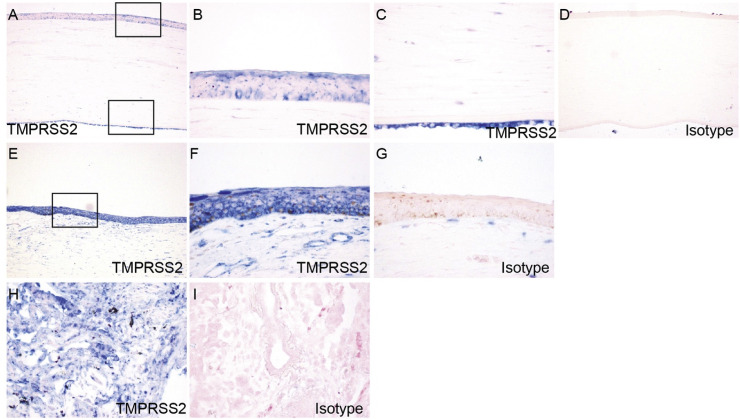
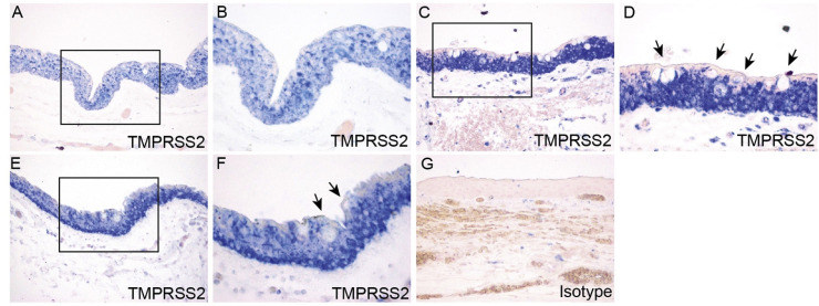
Results: Across all eye specimens, immunohistochemical analysis revealed expression of ACE2 in the conjunctiva, limbus, and cornea, with especially prominent staining in the superficial conjunctival and corneal epithelial surface. Surgical conjunctival specimens also showed expression of ACE2 in the conjunctival epithelium, especially prominent in the superficial epithelium, as well as weak or focal expression in the substantia propria. All eye and conjunctival specimens also expressed TMPRSS2. Finally, Western blot analysis of protein lysates from human corneal epithelium obtained during refractive surgery confirmed expression of ACE2 and TMPRSS2.

Conclusions: Together, these results suggest that ocular surface cells including conjunctiva are susceptible to infection by SARS-CoV-2, and could therefore serve as a portal of entry as well as a reservoir for person-to-person transmission of this virus. This highlights the importance of safety practices including face masks and ocular contact precautions in preventing the spread of COVID-19 disease.

Keywords: ACE2; COVID-19; Conjunctiva; Cornea; Human; Ocular surface; SARS-CoV-2.


Conflict of interest statement

The authors declare no conflicts of interest.

Figures

Fig. 1
Fig. 1
ACE2 and TMPRSS2 protein expression in human corneal epithelium. Tissue lysates were prepared from corneal epithelial cell samples collected from two patients and analyzed by Western blot, using antibodies against ACE2 or TMPRSS2. Beta-actin was used as the loading control.
Fig. 2
Fig. 2
Expression and localization of ACE2 in cornea and limbus. Post-mortem human eye sections were processed for immunohistochemistry with rabbit anti-ACE2 antibody. ACE2 expression was analyzed in cornea (A–C), limbus (E–F), and lung (H–I). Boxed region in (A) is shown at higher magnification in (B, C). Boxed region in (E) is shown at higher magnification in (F). Lung epithelium and vascular endothelium are indicated by red and blue arrow respectively. Immunohistochemistry with isotype- and concentration-matched normal IgG control was performed (D, G, J).
Fig. 3
Fig. 3
Expression and localization of ACE2 in conjunctiva in post-mortem globe. Paraffin-embedded human eye sections were processed for immunohistochemistry with rabbit anti-ACE2 antibody. ACE2 expression was analyzed in conjunctiva. Conjunctiva from two separate globes are depicted (A-B, C-D). For each individual globe, boxed region is shown at higher magnification. Arrows indicate vascular endothelium (A) and goblet cells (D). Immunohistochemistry with isotype- and concentration-matched normal IgG control was performed (E).
Fig. 4
Fig. 4
Expression and localization of ACE2 in surgical conjunctival specimens. Paraffin-embedded human surgical conjunctival sections were analyzed by immunohistochemistry with rabbit anti-ACE2 antibody. Boxed regions in (A) are shown at higher magnification in (B) and (C). Boxed region in (D) is shown at higher magnification in (E). Arrows indicate goblet cells (C, E). Immunohistochemistry with isotype- and concentration-matched normal IgG control was performed (F).
Fig. 5
Fig. 5
Expression and localization of TMPRSS2 in cornea and limbus. Paraffin-embedded human eye sections were processed for immunohistochemistry with rabbit anti-TMPRSS2 antibody. ACE2 expression was analyzed in cornea (A–C), limbus (E–F), and lung (H). Boxed region in (A) is shown at higher magnification in (B, C). Boxed region in (E) is shown at higher magnification in (F). Immunohistochemistry with isotype- and concentration-matched normal IgG control was performed (D, G, I).
Fig. 6
Fig. 6
Expression and localization of TMPRSS2 in conjunctiva in post-mortem globe. Paraffin-embedded human eye sections were processed for immunohistochemistry with rabbit anti-TMPRSS2 antibody. ACE2 expression was analyzed in conjunctiva. Conjunctiva from three separate globes are depicted (A-B, C-D, E-F). For each individual globe, boxed region is shown at higher magnification. Arrows indicate goblet cells (D, F). Immunohistochemistry with isotype- and concentration-matched normal IgG control was performed (G).
Fig. 7
Fig. 7
Expression and localization of TMPRSS2 in surgical conjunctival specimens. Paraffin-embedded human surgical conjunctival sections were analyzed by immunohistochemistry with rabbit anti-TMPRSS2 antibody. Boxed region in (A) is shown at higher magnification in (B). Arrows indicate goblet cells (B). Immunohistochemistry with isotype- and concentration-matched normal IgG control was performed (C).

Update of

Comment in

References

    1. Wu P., Duan F., Luo C., Liu Q., Qu X., Liang L. Characteristics of ocular findings of patients with coronavirus disease 2019 (COVID-19) in Hubei Province, China. JAMA Ophthalmol. 2020;138(5):575–578. doi: 10.1001/jamaophthalmol.2020.1291. - DOI - PMC - PubMed
    1. Seah I.Y.J., Anderson D.E., Kang A.E.Z., Wang L., Rao P., Young B.E. Assessing viral shedding and infectivity of tears in coronavirus disease 2019 (COVID-19) patients. Ophthalmology. 2020 doi: 10.1016/j.ophtha.2020.03.026. online ahead of print. - DOI - PMC - PubMed
    1. Zhang X., Chen X., Chen L., Deng C., Zou X., Liu W. The evidence of SARS-CoV-2 infection on ocular surface. Ocul Surf. 2020;18(3):360–362. doi: 10.1016/j.jtos.2020.03.010. - DOI - PMC - PubMed
    1. Jun I.S.Y., Hui K.K.O., Songbo P.Z. Perspectives on coronavirus disease 2019 control measures for ophthalmology clinics based on a Singapore center experience. JAMA Ophthalmol. 2020 doi: 10.1001/jamaophthalmol.2020.1288. - DOI - PubMed
    1. Napoli P.E., Nioi M., d’Aloja E., Fossarello M. The ocular surface and the coronavirus disease 2019: does a dual ‘ocular route’ exist? J Clin Med. 2020;9(5):E1269. doi: 10.3390/jcm9051269. online ahead of print. - DOI - PMC - PubMed

Publication types