Review
. 2020 Jun 11;21(11):4184.
doi: 10.3390/ijms21114184.

NLRP3 Inflammasome Activation in Adipose Tissues and Its Implications on Metabolic Diseases

Affiliations
Review

NLRP3 Inflammasome Activation in Adipose Tissues and Its Implications on Metabolic Diseases

Kelvin Ka-Lok Wu et al. Int J Mol Sci. .

Abstract

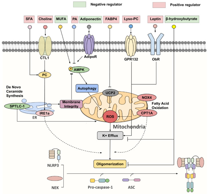
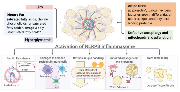
Adipose tissue is an active endocrine and immune organ that controls systemic immunometabolism via multiple pathways. Diverse immune cell populations reside in adipose tissue, and their composition and immune responses vary with nutritional and environmental conditions. Adipose tissue dysfunction, characterized by sterile low-grade chronic inflammation and excessive immune cell infiltration, is a hallmark of obesity, as well as an important link to cardiometabolic diseases. Amongst the pro-inflammatory factors secreted by the dysfunctional adipose tissue, interleukin (IL)-1β, induced by the NLR family pyrin domain-containing 3 (NLRP3) inflammasome, not only impairs peripheral insulin sensitivity, but it also interferes with the endocrine and immune functions of adipose tissue in a paracrine manner. Human studies indicated that NLRP3 activity in adipose tissues positively correlates with obesity and its metabolic complications, and treatment with the IL-1β antibody improves glycaemia control in type 2 diabetic patients. In mouse models, genetic or pharmacological inhibition of NLRP3 activation pathways or IL-1β prevents adipose tissue dysfunction, including inflammation, fibrosis, defective lipid handling and adipogenesis, which in turn alleviates obesity and its related metabolic disorders. In this review, we summarize both the negative and positive regulators of NLRP3 inflammasome activation, and its pathophysiological consequences on immunometabolism. We also discuss the potential therapeutic approaches to targeting adipose tissue inflammasome for the treatment of obesity and its related metabolic disorders.

Keywords: IL-1β; NLRP3 inflammasome; adipose tissue; immunometabolism; insulin sensitivity; metabolic disease; obesity.


Conflict of interest statement

The authors declare no conflict of interest. The funders had no role in the design of the study; in the collection, analyses, or interpretation of data; in the writing of the manuscript, or in the decision to publish the results.

Figures

Figure 1
Figure 1
Classical pathways for NLRP3 inflammasome activation. Upon stimulation of TLR4, IL-1R or TNFR, TNF receptor-associated factor 2 (TRAF2) and TNF receptor-associated factor 6 (TRAF6) recruit the inhibitor of nuclear factor-κB kinase α/β (IKKα/β) that drives the translocation of NF-κB subunits to the nucleus. This upregulates the transcription of NLRP3 and pro-IL-1β, which enables the following assembly of NLFPR3 inflammasome initiated by various PAMPs and DAMPs. Once activated, the dormant procaspase-1 is cleaved into active caspase-1, which initiates the processing of gasdermin D, pro-IL-1β and pro-IL-18 to their biologically active forms.
Figure 2
Figure 2
Key negative and positive regulators for NLRP3 inflammasome. Under nutrient overload, SFAs [such as palmitic acid (PA)] and choline are extensively incorporated into phosphatidylcholine (PC), which activates inositol-requiring enzyme 1α (IRE1α), whose endonuclease activity promotes NLPR3 inflammasome activation via an undefined mechanism. Furthermore, PC synthesis through the choline pathway reciprocally regulates the AMP-activated protein kinase (AMPK)–autophagy–ROS signaling axis by maintaining mitochondrial membrane integrity. On the other hand, monounsaturated fatty acids (MUFA) and adiponectin were identified as initiators of AMPK-dependent autophagy, that attenuate ROS production and K+ efflux, thereby suppressing NLRP3 activation. FABP4, lyso-PC, leptin and serine palmitoyltransferase long chain base subunit 1 (SPTLC-1), a key enzyme involved in de novo ceramide synthesis, all partake in NLRP3 inflammasome activation via increasing ROS production. NADPH oxidase 4 (NOX4) enhances the protein expression of carnitine palmitoyl-transferase 1A (CPT1A), a rate-limiting fatty acid oxidation-related enzyme, which is responsible for heightening NLRP3 inflammasome response through a largely unknown pathway. β-hydroxybutyrate (BHB) was unveiled as a potent NLRP3 inflammasome inhibitor, targeting both K+ efflux and ASC oligomerization.
Figure 3
Figure 3
Overview of NLRP3 inflammasome-associated metabolic consequences. (Image created with BioRender.com). * Activation of NLRP3 inflammasome by diverse metabolic stimuli (such as LPS, adipokines, hyperglycemia and mitochondrial dysfunction) leads to multiple metabolic and immune dysregulations including insulin resistance, altered immune cell composition, defective lipid handing and adipogenesis and increased fibrosis in white and brown fat depots. Detailed description and explanation for each consequence can be found in Section 3.

References

    1. Ouchi N., Parker J.L., Lugus J.J., Walsh K. Adipokines in inflammation and metabolic disease. Nat. Rev. Immunol. 2011;11:85–97. doi: 10.1038/nri2921. - DOI - PMC - PubMed
    1. Francisco V., Pino J., Gonzalez-Gay M.A., Mera A., Lago F., Gomez R., Mobasheri A., Gualillo O. Adipokines and inflammation: Is it a question of weight? Br. J. Pharmacol. 2018;175:1569–1579. doi: 10.1111/bph.14181. - DOI - PMC - PubMed
    1. Ferrante A.W., Jr. The immune cells in adipose tissue. Diabetes Obes. Metab. 2013;15(Suppl. 3):34–38. doi: 10.1111/dom.12154. - DOI - PMC - PubMed
    1. Rosen E.D., Spiegelman B.M. What we talk about when we talk about fat. Cell. 2014;156:20–44. doi: 10.1016/j.cell.2013.12.012. - DOI - PMC - PubMed
    1. Ikeda K., Maretich P., Kajimura S. The Common and Distinct Features of Brown and Beige Adipocytes. Trends Endocrinol. Metab. 2018;29:191–200. doi: 10.1016/j.tem.2018.01.001. - DOI - PMC - PubMed

MeSH terms