

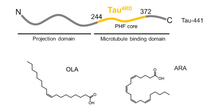
Unsaturated Fatty Acid-Induced Conformational Transitions and Aggregation of the Repeat Domain of Tau
- PMID: 32545360
- PMCID: PMC7321374
- DOI: 10.3390/molecules25112716
Unsaturated Fatty Acid-Induced Conformational Transitions and Aggregation of the Repeat Domain of Tau
Abstract
Background: The intrinsically disordered, amyloidogenic protein Tau associates with diverse classes of molecules, including proteins, nucleic acids, and lipids. Mounting evidence suggests that fatty acid molecules could play a role in the dysfunction of this protein, however, their interaction with Tau remains poorly characterized.
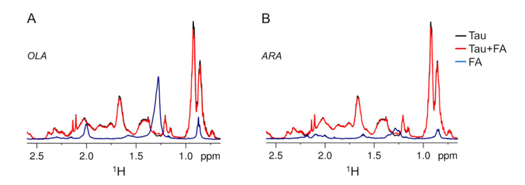
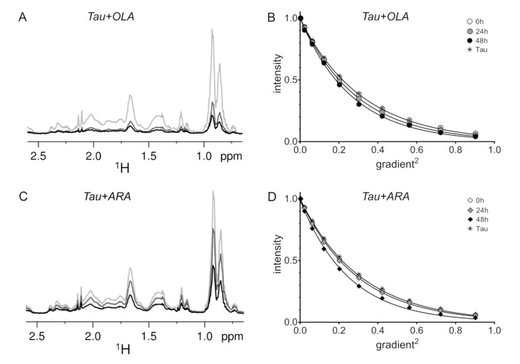
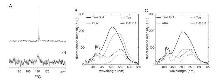
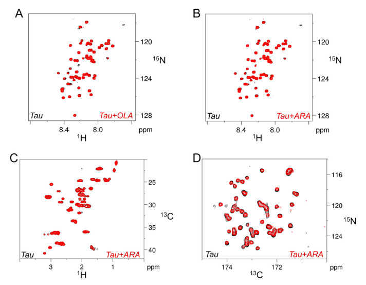
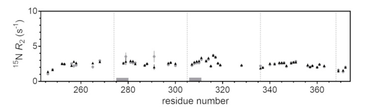
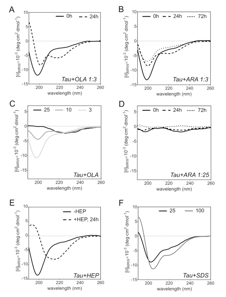
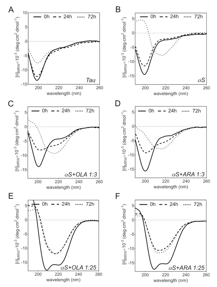
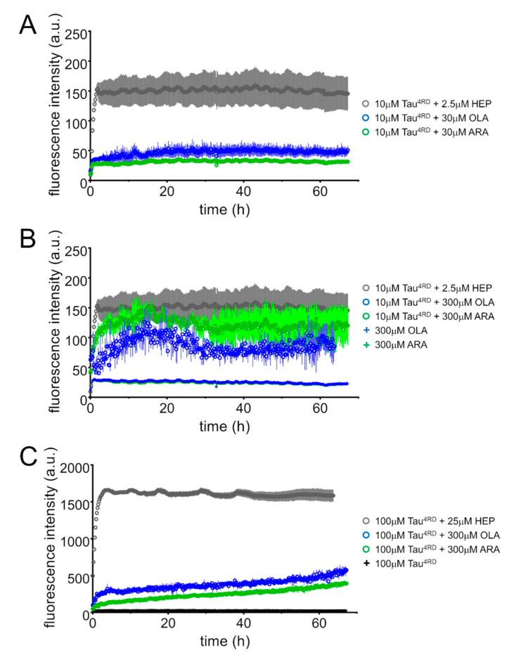
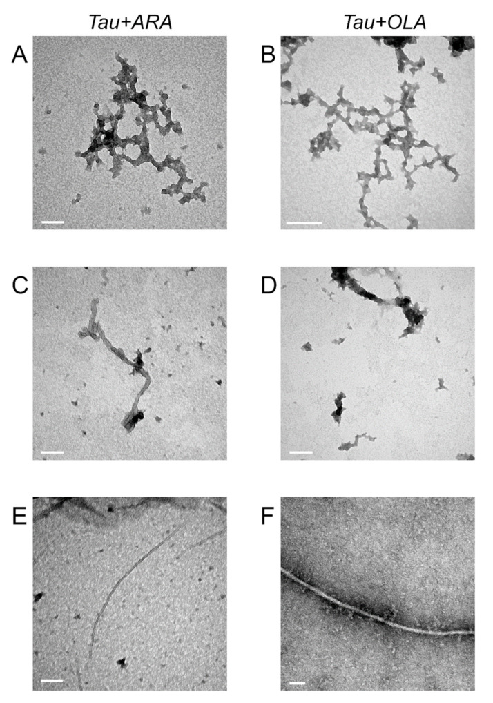
Methods: In a bid to elucidate the association of Tau with unsaturated fatty acids at the sub-molecular level, we carried out a variety of solution NMR experiments in combination with circular dichroism and fluorescence measurements. Our study shows that Tau4RD, the highly basic four-repeat domain of Tau, associates strongly with arachidonic and oleic acid assemblies in a high lipid/protein ratio, perturbing their supramolecular states and itself undergoing time-dependent structural adaptation. The structural signatures of Tau4RD/fatty acid aggregates appear similar for arachidonic acid and oleic acid, however, they are distinct from those of another prototypical intrinsically disordered protein, α-synuclein, when bound to these lipids, revealing protein-specific conformational adaptations. Both fatty acid molecules are found to invariably promote the self-aggregation of Tau4RD and of α-synuclein.
Conclusions: This study describes the reciprocal influence that Tau4RD and fatty acids exert on their conformational states, contributing to our understanding of fundamental aspects of Tau/lipid co-assembly.
Keywords: NMR spectroscopy; Tau; fatty acids; neurodegeneration; protein aggregation; protein–lipid interactions.
Conflict of interest statement
The authors declare no conflict of interest. The funders had no role in the design of the study; in the collection, analyses, or interpretation of data; in the writing of the manuscript, or in the decision to publish the results.
Figures
Similar articles
-
Tau Interacts with the C-Terminal Region of α-Synuclein, Promoting Formation of Toxic Aggregates with Distinct Molecular Conformations.Biochemistry. 2019 Jun 25;58(25):2814-2821. doi: 10.1021/acs.biochem.9b00215. Epub 2019 Jun 7. Biochemistry. 2019. PMID: 31132261 Free PMC article.
-
Synergistic Amyloid Switch Triggered by Early Heterotypic Oligomerization of Intrinsically Disordered α-Synuclein and Tau.J Mol Biol. 2018 Aug 3;430(16):2508-2520. doi: 10.1016/j.jmb.2018.04.020. Epub 2018 Apr 25. J Mol Biol. 2018. PMID: 29704492
-
α-Synuclein Fibrils Exhibit Gain of Toxic Function, Promoting Tau Aggregation and Inhibiting Microtubule Assembly.J Biol Chem. 2016 Jul 15;291(29):15046-56. doi: 10.1074/jbc.M116.736355. Epub 2016 May 19. J Biol Chem. 2016. PMID: 27226637 Free PMC article.
-
Solution NMR insights into dynamic supramolecular assemblies of disordered amyloidogenic proteins.Arch Biochem Biophys. 2020 Apr 15;683:108304. doi: 10.1016/j.abb.2020.108304. Epub 2020 Feb 22. Arch Biochem Biophys. 2020. PMID: 32097611 Review.
-
A flash in the pan: dissecting dynamic amyloid intermediates using fluorescence.FEBS Lett. 2013 Apr 17;587(8):1096-105. doi: 10.1016/j.febslet.2013.02.044. Epub 2013 Mar 1. FEBS Lett. 2013. PMID: 23458258 Free PMC article. Review.
Cited by
-
Lipid Dys-Homeostasis Contributes to APOE4-Associated AD Pathology.Cells. 2022 Nov 15;11(22):3616. doi: 10.3390/cells11223616. Cells. 2022. PMID: 36429044 Free PMC article.
-
Much More Than a Cytoskeletal Protein: Physiological and Pathological Functions of the Non-microtubule Binding Region of Tau.Front Neurol. 2020 Oct 19;11:590059. doi: 10.3389/fneur.2020.590059. eCollection 2020. Front Neurol. 2020. PMID: 33193056 Free PMC article. Review.
-
Lipid profiles in the cerebrospinal fluid of rats with 6-hydroxydopamine-induced lesions as a model of Parkinson's disease.Front Aging Neurosci. 2023 Jan 20;14:1077738. doi: 10.3389/fnagi.2022.1077738. eCollection 2022. Front Aging Neurosci. 2023. PMID: 36742201 Free PMC article.
-
Membrane charge primes the necroptotic kinase RIPK3 for amyloid assembly.Commun Chem. 2025 Aug 21;8(1):252. doi: 10.1038/s42004-025-01658-0. Commun Chem. 2025. PMID: 40841823 Free PMC article.
-
Espresso Coffee Mitigates the Aggregation and Condensation of Alzheimer's Associated Tau Protein.J Agric Food Chem. 2023 Aug 2;71(30):11429-11441. doi: 10.1021/acs.jafc.3c01072. Epub 2023 Jul 19. J Agric Food Chem. 2023. PMID: 37466260 Free PMC article.
References
-
- Schweers O., Schönbrunn-Hanebeck E., Marx A., Mandelkow E. Structural studies of tau protein and Alzheimer paired helical filaments show no evidence for beta-structure. J. Biol. Chem. 1994;269:24290–24297. - PubMed
MeSH terms
Substances
Grants and funding
LinkOut - more resources
Full Text Sources
