. 2020 Jun 11;21(11):4179.
doi: 10.3390/ijms21114179.

Apelin Controls Angiogenesis-Dependent Glioblastoma Growth

Affiliations

Apelin Controls Angiogenesis-Dependent Glioblastoma Growth

Anne Frisch et al. Int J Mol Sci. .

Abstract

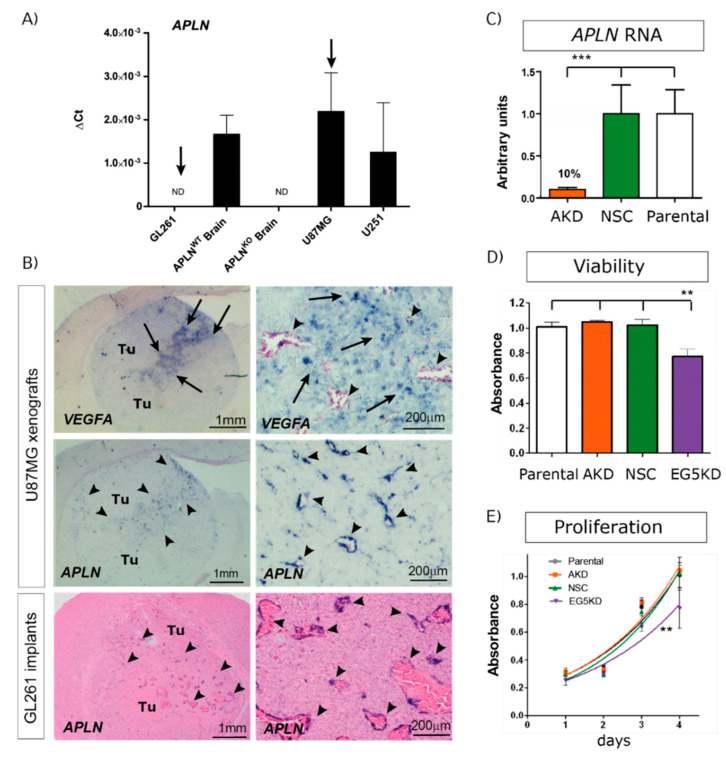
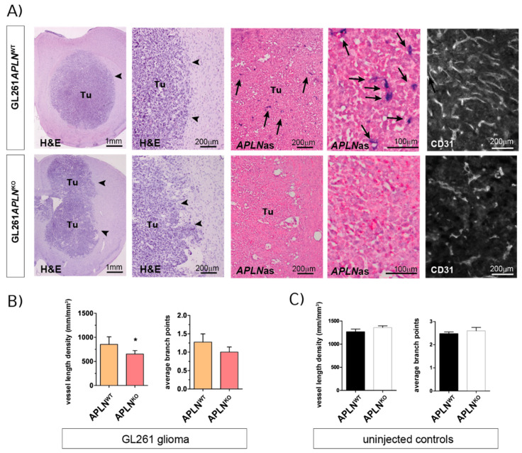
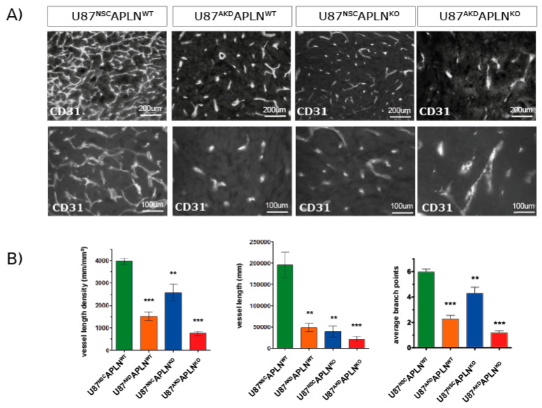
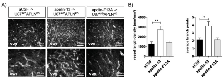
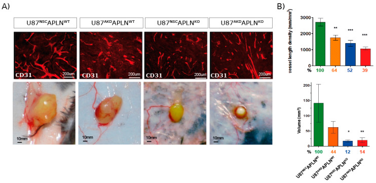
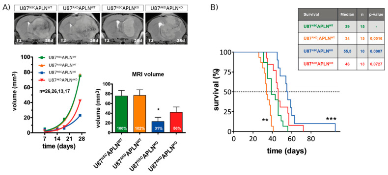
Glioblastoma (GBM) present with an abundant and aberrant tumor neo-vasculature. While rapid growth of solid tumors depends on the initiation of tumor angiogenesis, GBM also progress by infiltrative growth and vascular co-option. The angiogenic factor apelin (APLN) and its receptor (APLNR) are upregulated in GBM patient samples as compared to normal brain tissue. Here, we studied the role of apelin/APLNR signaling in GBM angiogenesis and growth. By functional analysis of apelin in orthotopic GBM mouse models, we found that apelin/APLNR signaling is required for in vivo tumor angiogenesis. Knockdown of tumor cell-derived APLN massively reduced the tumor vasculature. Additional loss of the apelin signal in endothelial tip cells using the APLN-knockout (KO) mouse led to a further reduction of GBM angiogenesis. Direct infusion of the bioactive peptide apelin-13 rescued the vascular loss-of-function phenotype specifically. In addition, APLN depletion massively reduced angiogenesis-dependent tumor growth. Consequently, survival of GBM-bearing mice was significantly increased when APLN expression was missing in the brain tumor microenvironment. Thus, we suggest that targeting vascular apelin may serve as an alternative strategy for anti-angiogenesis in GBM.

Keywords: APLN; APLNR; Apelin-13; GBM angiogenesis; glioblastoma.


Conflict of interest statement

The authors declare no conflict of interest.

Figures

Figure 1
Figure 1
Angiogenic factor apelin (APLN) expression in glioblastoma (GBM) cells and the tumor neo-vasculature. (A) Expression analysis of APLN RNA was performed by qPCR on the human cells U87MG and U251, the murine GL261 cells and on APLNWT or APLNKO mouse brain tissue. APLN RNA expression was not detectable (n.d.) in GL261 cells but high in U87MG cells. (B) In situ hybridization against mouse APLN showed no expression in implanted mouse GL261 tumor cells but an upregulation of APLN in the tumor vessels of GL261 or U87MG gliomas (arrowheads). The right panel is the magnifications within the tumor that is depicted in the overview panel on the left. Note that vascular APLN RNA expression is highest in hypoxic regions marked by human vascular endothelial growth factor (VEGFA) RNA expression (arrows) in the U87MG xenografts. (C) Lentiviral transduction of U87MG parental cells caused shRNAmir-mediated stable APLN knock-down (AKD) by 90% (as analyzed by qPCR) compared to the non-silencing shRNA control (NSC) transduced cells. (D) Cell viability, as well as in vitro proliferation (E), was unchanged in U87AKD cells compared to U87NSC control or untransduced U87MG cells. In contrast, U87 cells transduced with the shRNA against the kinesin EG5 significantly reduced viability and proliferation of U87E5KD cells compared to U87NSC control. Data are obtained from more than 3 independent experiments each and reported as mean +/-SEM; statistical significance (one-way ANOVA plus Bonferroni’s post hoc test) is indicated ** p < 0.005, *** p < 0.0005. WT = wildtype; KO = knockout.
Figure 2
Figure 2
Endothelial APLN expression controls formation of a complex GBM vasculature. Orthotopic GL261 implants in APLNWT or APLNKO mice 21 days post-implantation (dpi). (A) The panels on the left indicate the tumor (tumor boarder highlighted by arrowheads) in the right brain hemisphere in overview and close up view on Hematoxylin & Eosin (H&E) sections. The in situ hybridization panels in the middle show the loss-of vascular APLN expression (arrows) in the tumor in overview and close up view when comparing APLNWT to APLNKO mice. The CD31-immunostaining shown in the panel on the right illustrates the reduced vessel density in APLNKO mice. (B) Vessel length density (VLD) was assessed on CD31 immunofluorescent brain slides and demonstrated a significant decrease in APLNKO mice. In addition, vascular complexity measured by the average branch points (ABP) was reduced in APLNKO as compared to APLNWT. (C) In the healthy brain, VLD and ABP do not differ in APLNKO compared to APLNWT mice. Data of n = 9 APLNWT vs. 6 APLNKO mice are reported as mean +/-SEM; statistical significance (students test) is indicated * p < 0.05.
Figure 3
Figure 3
GBM-and endothelial cell-derived APLN are both controlling sprouting angiogenesis. (A) U87MG was implanted into immunodeficient mice and grown to big xenografts within 28 dpi. Fluorescent immunostaining for CD31 was performed and is depicted in an overview and a close up view for every xenograft. (B) The microvasculature in the green fluorescent protein (GFP)-positive tumor area was analyzed by stereomorphology. In comparison to U87NSCAPLNWT controls (n = 8), VLD in U87AKDAPLNWT xenografts (n = 13) was reduced by 62% and vessel length by 75%. Additional loss-of-APLN expression in the tumor neo-vasculature of the APLNKO mouse (U87AKDAPLNKO, n = 10) lead to further reduction of the tumor vasculature. VLD was also reduced when only the tumor cells express APLN (U87NSCAPLNKO, n = 9) but was the highest of all manipulated xenografts. While vascular complexity measured by vascular branch points (ABP values are 6, 2.3; 4.3; and 1.2, respectively) reflects the results seen by VLD, the total vessel length was highly reduced in all three xenografts with reduction of APLN expression. Data are reported as mean +/−SEM; statistical significance (one-way ANOVA plus Bonferroni´s post hoc test) is indicated ** p < 0.005, *** p < 0.0005.
Figure 4
Figure 4
Apelin peptide rescues the vascular loss-of-function phenotype. (A) Intracerebral infusion of 30 µg of apelin-13 peptide (n = 9) increased glioma angiogenesis in U87AKDAPLNKO xenografts compared to infusion of artificial cerebrospinal fluid (aCSF, n = 8) or apelin-F13A (n = 6) antagonist as shown by von Willebrand factor (VWF) staining. (B) Quantification on a Stereoinvestigator resulted in a VLD of 1269 mm/mm3 in U87AKDAPLNKO xenografts infused with aCSF only; 2573 mm/mm3 with the APLN receptor (APLNR) agonist apelin-13 peptide or 1419 mm/mm3 with the APLNR antagonist apelin-F13A. ABP obtained were 2.2; 3.9 and 2.2, respectively. Data are reported as mean +/−SEM; statistical significance (one-way ANOVA plus Bonferroni´s post hoc test) is indicated. * p < 0.05, ** p < 0.005.
Figure 5
Figure 5
Apelin is required for compact tumor growth. Mice were inoculated subcutaneously with U87 cells and grown for 28 dpi. (A) Tumor volumes were measured, and vessel density was quantified on CD31-immunostained sections. Representative pictures of subcutaneous tumors are shown. (B) VLD and tumor volume was significantly attenuated upon reduction of APLN expression as compared to U87NSCAPLNWT controls (percentages compared to the control group are indicated). Tumor volumes were reduced to 50% in APLN knockdown U87AKDAPLNWT xenografts. Further reduction to less than 15% was observed in APLNKO mice. Number of cell implantations for U87NSCAPLNWT, U87AKDAPLNWT, U87NSCAPLNKO, U87AKDAPLNKO were n = 38, 31, 18, 24, while tumor take was 87%, 90% 72%, 50%, respectively. For VLD analysis n = 7, 6, 7, 6 of the respective xenografts were analyzed. Data are reported as mean +/−SEM; statistical significance (one-way ANOVA plus Bonferroni’s post hoc test) is indicated * p < 0.05, ** p < 0.005, *** p < 0.0005.
Figure 6
Figure 6
Loss-of-APLN in the tumor microenvironment increases survival of GBM mice. Human U87 cells modulated for APLN levels were orthotopically implanted into APLNWT or APLNKO mice. (A) T2-weighted magnetic resonance imaging (MRI) was performed weekly and MRI volumes were measured producing an in vivo growth curve for every xenograft. After 28 days, U87NSC or U87AKD cells in APLNKO mice showed reduced tumor volume by 69% and 44%, respectively, compared to U87NSCAPLNWT controls. Number of mice per group are indicated. Data are reported as mean +/−SEM; statistical significance (one-way ANOVA plus Bonferroni’s post hoc test) is indicated * p < 0.05. (B) A mouse survival experiment with orthotopically implanted tumor cells was performed and mice sacrificed at humane endpoints. Median survival differed significantly in U87AKDAPLNWT (shorter survival) and U87NSCAPLNKO (longer survival) mice compared to U87NSCAPLNWT control mice. U87AKD xenografts in APLNKO mice showed a trend to longer survival. Number of mice per group are indicated. Survival data are shown as Kaplan-Meier Curves and significant differences between the experimental groups and the U87NSCAPLNWT control group assessed by long-rank (Mantel-Cox) test is given in the table and is indicated in the graph by * p < 0.05, ** p < 0.01, *** p < 0.001.

References

    1. Kleihues P., Cavenee W.K. Pathology and Genetics of Tumours of the Nervous System. IARC Press; Lyon, France: 2000.
    1. Stupp R., Mason W.P., van den Bent M.J., Weller M., Fisher B., Taphoorn M.J., Belanger K., Brandes A.A., Marosi C., Bogdahn U., et al. Radiotherapy plus concomitant and adjuvant temozolomide for glioblastoma. N. Engl. J. Med. 2005;352:987–996. doi: 10.1056/NEJMoa043330. - DOI - PubMed
    1. Weller M., van den Bent M., Tonn J.C., Stupp R., Preusser M., Cohen-Jonathan-Moyal E., Henriksson R., Le Rhun E., Balana C., Chinot O., et al. European Association for Neuro-Oncology (EANO) guideline on the diagnosis and treatment of adult astrocytic and oligodendroglial gliomas. Lancet Oncol. 2017;18:e315–e329. doi: 10.1016/S1470-2045(17)30194-8. - DOI - PubMed
    1. Louis D.N., Perry A., Reifenberger G., von Deimling A., Figarella-Branger D., Cavenee W.K., Ohgaki H., Wiestler O.D., Kleihues P., Ellison D.W., et al. The 2016 World Health Organization Classification of Tumors of the Central Nervous System: A summary. Acta Neuropathol. 2016;131:803–820. doi: 10.1007/s00401-016-1545-1. - DOI - PubMed
    1. Hegi M.E., Diserens A.C., Gorlia T., Hamou M.F., de Tribolet N., Weller M., Kros J.M., Hainfellner J.A., Mason W., Mariani L., et al. MGMT gene silencing and benefit from temozolomide in glioblastoma. N. Engl. J. Med. 2005;352:997–1003. doi: 10.1056/NEJMoa043331. - DOI - PubMed

MeSH terms