. 2020 Jun 11;12(6):1544.
doi: 10.3390/cancers12061544.

Epidemiology of Signet Ring Cell Adenocarcinomas

Affiliations

Epidemiology of Signet Ring Cell Adenocarcinomas

Matthew G K Benesch et al. Cancers (Basel). .

Abstract

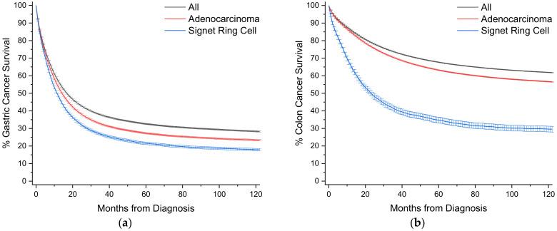
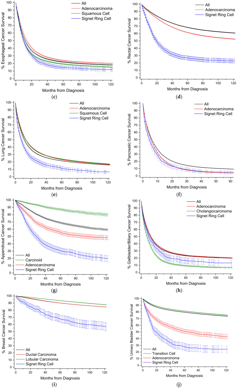
Signet ring cell adenocarcinomas (SRCCs) are a rare histological subtype of adenocarcinomas with a poor prognosis, typically due to advanced disease at diagnosis. A signet ring cell, mimicking its moniker, contains abundant intracytoplasmic mucin that pushes the nucleus to the periphery. In these cancers, this cell feature comprises more than 50% of the tumor. Despite predilection for the gastrointestinal tract, and in particular the stomach, primary SRCCs are also described in other sites, typically in case reports. This literature, however, lacks a standardized overview of the SRCC disease entity. Using a retrospective cohort approach, we summarize the clinicodemographic and mortality outcomes of SRCCs in thirteen primary sites, comprising 95% of all SRCCs in the Surveillance, Epidemiology, and End Results Program (SEER), a population-level cancer database covering nearly one-third of the United States population. SRCCs general trends compared to matching nonvariant adenocarcinomas are earlier age of onset, with initial presentation favoring higher rates of regional or distant disease presentation and poor tumor differentiation. After multivariable analysis, SRCCs typically have worse overall survivals, but substantial variances exist depending on tumor location. Identifying SRCCs at earlier disease stages is likely the single most important intervention to improving outcomes for these patients.

Keywords: CDH1; E-cadherin; cancer; chemotherapy; diffuse type; histopathology; radiotherapy; surgery.


Conflict of interest statement

The authors declare no conflict of interest.

Figures

Figure 1
Figure 1
(a) Representative histological slide of poorly differentiated conventional gastric adenocarcinoma. (b) Representative histological slide of gastric signet adenocarcinoma, illustrating mucin-filled cytoplasm with nucleus pushed to the periphery. Figures sourced from Wikimedia Commons, public domain [6,7].
Figure 2
Figure 2
(a) Distribution of signet ring cell tumors in SEER, 1975–2016, total of 41,847 cases. (b) Distribution of all solid (non-blood borne), non-signet ring cell tumors in SEER, 1975–2016, total of 9.56 million cases. In both plots data labels are percentages. Markers omitted if less than 1%.
Figure 3
Figure 3
Kaplan-Meier survival curves. All survivor functions are shown with 95% confidence intervals. (a) Gastric cancer. (b) Colon cancer. (c) Esophageal cancer. (d) Rectal cancer. (e) Lung cancer. (f) Pancreatic cancer. (g) Appendiceal cancer. (h) Gallbladder/Biliary cancer. (i) Breast cancer. (j) Urinary Bladder cancer. (k) Small Bowel cancer. (l) Ovarian cancer. (m) Prostate cancer. In these curves, “All” represents the curves for all cancers within that site, with subtypes shown as labelled.
Figure 3
Figure 3
Kaplan-Meier survival curves. All survivor functions are shown with 95% confidence intervals. (a) Gastric cancer. (b) Colon cancer. (c) Esophageal cancer. (d) Rectal cancer. (e) Lung cancer. (f) Pancreatic cancer. (g) Appendiceal cancer. (h) Gallbladder/Biliary cancer. (i) Breast cancer. (j) Urinary Bladder cancer. (k) Small Bowel cancer. (l) Ovarian cancer. (m) Prostate cancer. In these curves, “All” represents the curves for all cancers within that site, with subtypes shown as labelled.

References

    1. Laufman H., Saphir O. Primary linitis plastica type of carcinoma of the colon. AMA Arch. Surg. 1951;62:79–91. doi: 10.1001/archsurg.1951.01250030082009. - DOI - PubMed
    1. Saphir O. Signet-ring cell carcinoma of the urinary bladder. Am. J. Pathol. 1955;31:223–231. - PMC - PubMed
    1. El Hussein S., Khader S.N. Primary signet ring cell carcinoma of the pancreas: Cytopathology review of a rare entity. Diagn. Cytopathol. 2019;47:1314–1320. doi: 10.1002/dc.24324. - DOI - PubMed
    1. Bosman F.T., Carneiro F., Hruban R.H., Theise N.D. WHO Classification of Tumours of the Digestive System. World Health Organization; Geneva, Switzerland: 2010.
    1. WHO . Digestive System Tumours, WHO Classification of Tumours Series. 5th ed. International Agency for Research on Cancer; Lyon, France: 2019.

LinkOut - more resources