. 2020 Jun 11;21(11):4190.
doi: 10.3390/ijms21114190.

RNA Binding Proteins as Drivers and Therapeutic Target Candidates in Pancreatic Ductal Adenocarcinoma

Affiliations

RNA Binding Proteins as Drivers and Therapeutic Target Candidates in Pancreatic Ductal Adenocarcinoma

Markus Glaß et al. Int J Mol Sci. .

Abstract

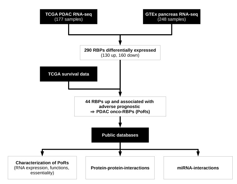
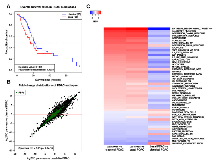
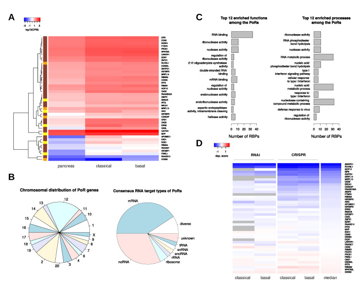
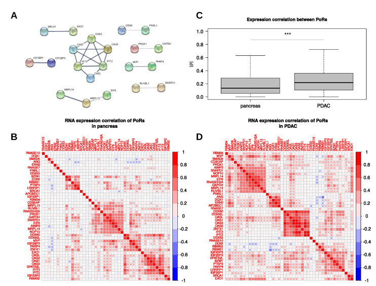
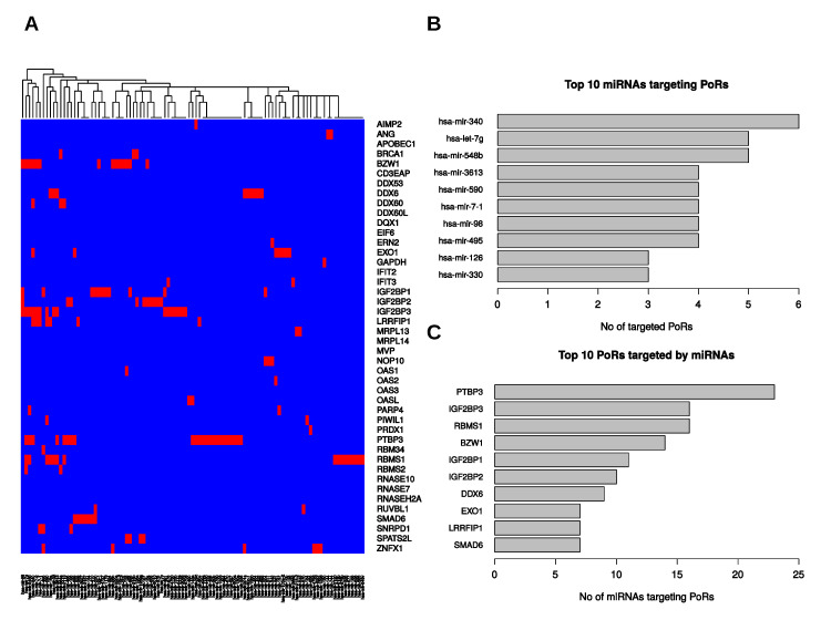
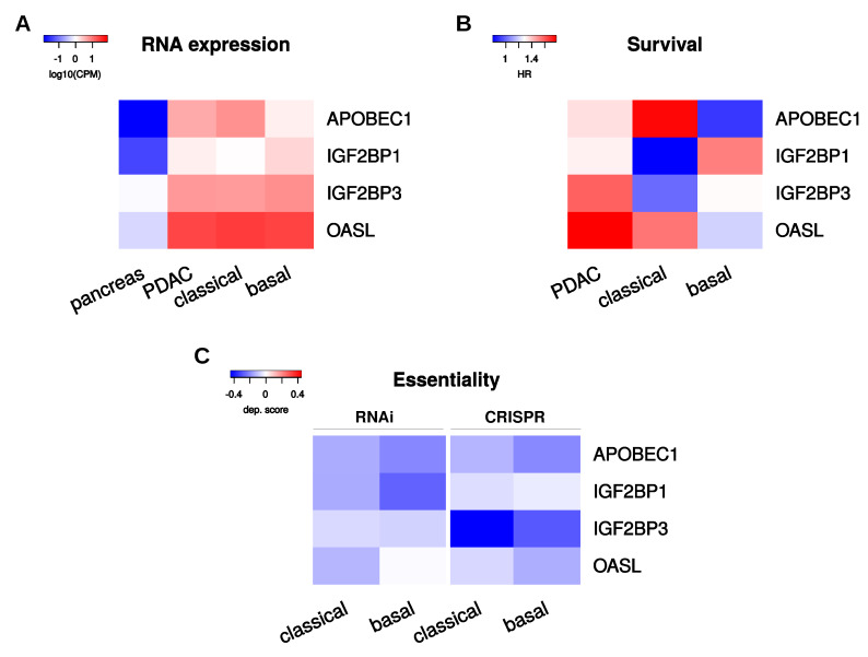
Pancreatic ductal adenocarcinomas (PDAC) belong to the most frequent and most deadly malignancies in the western world. Mutations in KRAS and TP53 along with some other frequent polymorphisms occur almost universally and are likely to be responsible for tumor initiation. However, these mutations cannot explain the heterogeneity in therapeutic responses observed in PDAC patients, which limits efficiency of current therapeutic strategies. Instead, recent classifications of PDAC tumor samples are based on transcriptomics data and thus include information about epigenetic, transcriptomic, and post-transcriptomic deregulations. RNA binding proteins (RBPs) are important post-transcriptional regulators involved in every aspect of the RNA life cycle and thus considerably influence the transcriptome. In this study, we systematically investigated deregulated expression, prognostic value, and essentiality reported for RBPs in PDAC or PDAC cancer models using publicly available data. We identified 44 RBPs with suggested oncogenic potential. These include various proteins, e.g., IGF2 mRNA binding proteins (IGF2BPs), with reported tumor-promoting roles. We further characterized these RBPs and found common patterns regarding their expression, interaction, and regulation by microRNAs. These analyses suggest four prime candidate oncogenic RBPs with partially validated target potential: APOBEC1, IGF2BP1 and 3, and OASL.

Keywords: APOBEC1; IGF2BP1; IGF2BP3; OASL; RNA binding proteins (RBPs); pancreatic ductal adenocarcinoma (PDAC).


Conflict of interest statement

The authors declare no conflict of interest.

Figures

Figure 1
Figure 1
Analysis of pancreatic ductal adenocarcinomas (PDAC) onco-RNA binding proteins (RBPs). Flowchart depicting the analysis pipeline for the in silico identification and characterization of RBPs with oncogenic characteristics in PDAC using public data sets. Black boxes represent data sources, white boxes denote analysis steps.
Figure 2
Figure 2
PDAC subtype properties. (A) Kaplan–Meier-curves showing overall survival rates for classical (blue) and basal-like (red) PDAC. (B) RNA expression changes of protein-coding genes in classical and basal-like PDAC subtypes compared to normal pancreas samples. Green points mark RNA binding proteins (RBPs). (C) Heatmap showing normalized enrichment scores (NES) obtained from gene set enrichment analyses (GSEA) of the 50 MSigDB hallmark gene sets using fold changes obtained by the comparisons of pancreas against classical PDAC (first column), pancreas against basal-like PDAC (second column), and basal-like against classical PDAC (third column).
Figure 3
Figure 3
Properties of PDAC onco-RBPs (PoRs). (A) Average normalized RNA expression values (log10 CPM) of the 44 PoRs in pancreas as well as in classical and in basal-like PDAC samples. The color bar on the left side encodes subtype specificity of oncogene properties (yellow—upregulated and adverse prognostic only in classical PDAC; orange—upregulated and adverse prognostic only in basal-like PDAC; brown—upregulated and adverse prognostic in both PDAC subtypes). (B) Chromosomal distribution (left) and consensus RNA target types (right) of the PoRs. (C) Top 12 significantly (FDR < 0.05) enriched functions (left) and biological processes (right) among the PoRs. Gene Ontology terms are sorted according to the significance of their enrichment. (D) Average dependency scores of the PoRs in PDAC subtype specific cell lines obtained by knockdown via RNAi or CRISPR knockout. Gray color denotes missing values.
Figure 4
Figure 4
PoR interactions. (A) Known physical interactions between PoRs according to the STRING database. (B,D) Spearman’s correlation coefficients of the PoR RNA expression in pancreas (B) and PDAC (D) RNA-seq samples. (C) Distribution of Spearman’s correlation coefficient magnitudes (|ρ|) obtained from comparisons among the 44 PoRs in pancreas and PDAC RNA-seq samples. ***: Mann–Whitney test p-value < 0.001.
Figure 5
Figure 5
Putative interactions between PoRs and microRNAs. (A) Predicted PoR–miRNA-interactions that were associated with significant (p < 0.05) negative expression correlation (Spearman). Red color encodes inferred interaction, blue means no interaction. (B) Top 10 miRNAs targeting the most PoRs. (C) Top 10 PoRs targeted by the most miRNAs.
Figure 6
Figure 6
Selected PoRs. (A) Average normalized RNA expression values (log10 CPM) of the four selected PoRs in pancreas and PDAC samples comprised of the whole (PDAC) tumor sample set as well as subtype specific subsets. (B) Hazard ratios (HR) of the four selected PoRs determined from overall survival rates between low and high RNA expression. (C) Average dependency scores of the four selected PoRs obtained from RNAi-derived depletion as well as CRISPR-derived deletion in classical and basal-like PDAC derived cell lines.

References

    1. Bailey P., Chang D.K., Nones K., Johns A.L., Patch A.M., Gingras M.C., Miller D.K., Christ A.N., Bruxner T.J., Quinn M.C., et al. Genomic analyses identify molecular subtypes of pancreatic cancer. Nature. 2016;531:47–52. doi: 10.1038/nature16965. - DOI - PubMed
    1. Rahib L., Smith B.D., Aizenberg R., Rosenzweig A.B., Fleshman J.M., Matrisian L.M. Projecting cancer incidence and deaths to 2030: The unexpected burden of thyroid, liver, and pancreas cancers in the United States. Cancer Res. 2014;74:2913–2921. doi: 10.1158/0008-5472.CAN-14-0155. - DOI - PubMed
    1. Chen C.C., Chen L.L., Li C.P., Hsu Y.T., Jiang S.S., Fan C.S., Chua K.V., Huang S.X., Shyr Y.M., Chen L.T., et al. Myeloid-derived macrophages and secreted HSP90α induce pancreatic ductal adenocarcinoma development. Oncoimmunology. 2018;7:e1424612. doi: 10.1080/2162402X.2018.1424612. - DOI - PMC - PubMed
    1. Knudsen E.S., Balaji U., Mannakee B., Vail P., Eslinger C., Moxom C., Mansour J., Witkiewicz A.K. Pancreatic cancer cell lines as patient-derived avatars: Genetic characterisation and functional utility. Gut. 2018;67:508–520. doi: 10.1136/gutjnl-2016-313133. - DOI - PMC - PubMed
    1. Kamisawa T., Wood L.D., Itoi T., Takaori K. Pancreatic cancer. Lancet. 2016;388:73–85. doi: 10.1016/S0140-6736(16)00141-0. - DOI - PubMed

MeSH terms