Review
. 2020 Jun 12;21(12):4193.
doi: 10.3390/ijms21124193.

Computer-Aided Ligand Discovery for Estrogen Receptor Alpha

Affiliations
Review

Computer-Aided Ligand Discovery for Estrogen Receptor Alpha

Divya Bafna et al. Int J Mol Sci. .

Abstract

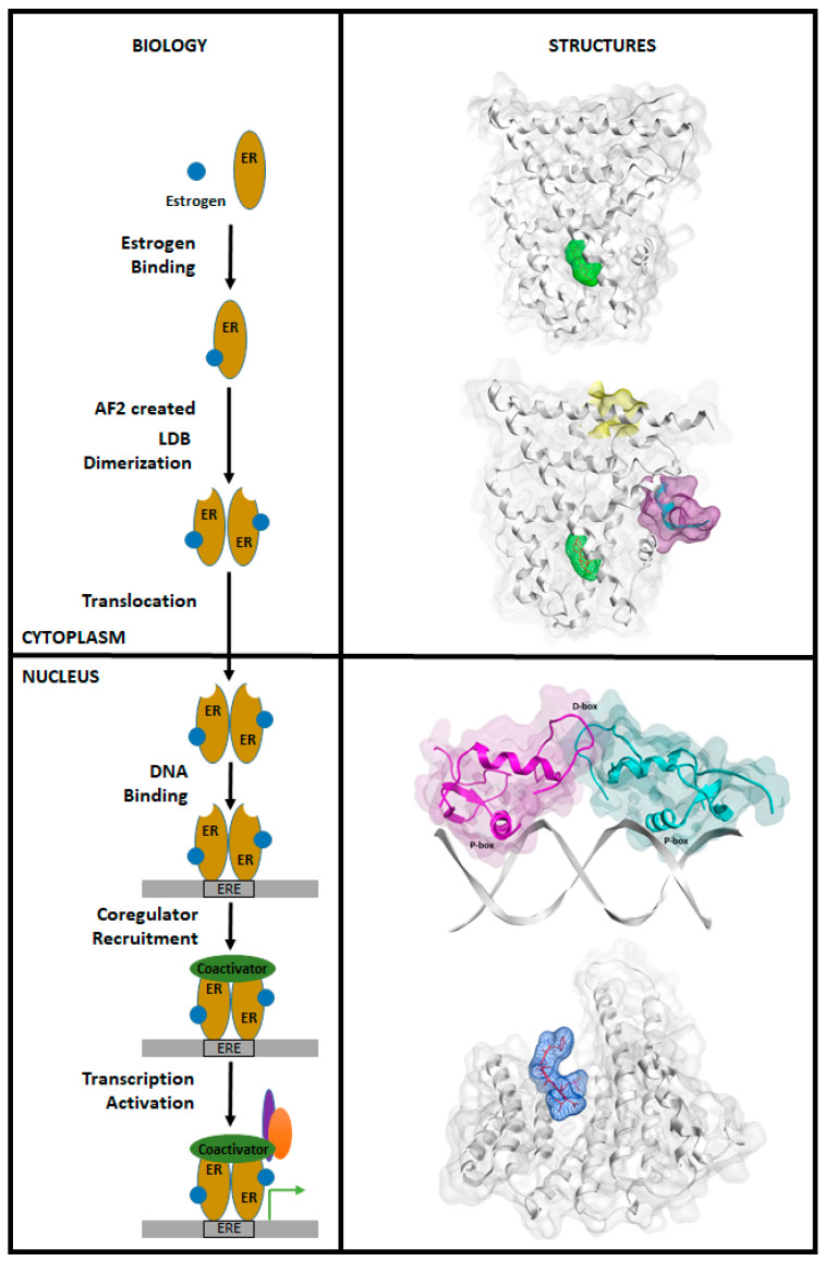
Breast cancer (BCa) is one of the most predominantly diagnosed cancers in women. Notably, 70% of BCa diagnoses are Estrogen Receptor α positive (ERα+) making it a critical therapeutic target. With that, the two subtypes of ER, ERα and ERβ, have contrasting effects on BCa cells. While ERα promotes cancerous activities, ERβ isoform exhibits inhibitory effects on the same. ER-directed small molecule drug discovery for BCa has provided the FDA approved drugs tamoxifen, toremifene, raloxifene and fulvestrant that all bind to the estrogen binding site of the receptor. These ER-directed inhibitors are non-selective in nature and may eventually induce resistance in BCa cells as well as increase the risk of endometrial cancer development. Thus, there is an urgent need to develop novel drugs with alternative ERα targeting mechanisms that can overcome the limitations of conventional anti-ERα therapies. Several functional sites on ERα, such as Activation Function-2 (AF2), DNA binding domain (DBD), and F-domain, have been recently considered as potential targets in the context of drug research and discovery. In this review, we summarize methods of computer-aided drug design (CADD) that have been employed to analyze and explore potential targetable sites on ERα, discuss recent advancement of ERα inhibitor development, and highlight the potential opportunities and challenges of future ERα-directed drug discovery.

Keywords: breast cancer; computer-aided drug design; estrogen receptor; virtual screening.


Conflict of interest statement

The authors declare no conflict of interest.

Figures

Figure 1
Figure 1
(a) Evolution of BCa treatment and therapy; and (b) Computer-aided drug design techniques for standard drug discovery pipeline.
Figure 1
Figure 1
(a) Evolution of BCa treatment and therapy; and (b) Computer-aided drug design techniques for standard drug discovery pipeline.
Figure 2
Figure 2
(a) ERα and ERβ domain organization; (b) homology of ERα and ERβ at different domains; and (c) F-domain sequence alignment between ERα and ERβ.
Figure 3
Figure 3
E2 (Green) bound to wild-type ERα at the EBS (pdb: 1QKU). The hydrogen bonds are depicted using dashed lines (orange). E2 forms two hydrogen bonds on one side with Arg394 and Glu353 and forms one on the other side with His524.
Figure 4
Figure 4
The biology of classic ER transcriptional mechanism (left) with the corresponding structures (right): the first structure depicts E2 (green) bound to the ER-LBD; the second structure depicts an E2 (green) activated ER with an coactivator docked to the created AF-2 site (purple) along with the potential dynamic BF3 site (yellow); the third structure shows a translocated DBD/DBD ER dimer bound to DNA; and the fourth structure shows the F-domain bound to an AF-1 mediated transcription repressor protein, 14-3-3 (white).
Figure 5
Figure 5
(a) E2 (green) bound to ERα (white) and the coactivator peptide (yellow) (pdb: 3UUD) with the helix numbers from 1 to 12 specified. The binding of E2 moves the H12 to its active conformation, creating the AF2 site that binds to Co-activator proteins. (b) OHT (red) bound ERα (white) (pdb: 3ERT). Binding of OHT moves the H12 to an antagonistic conformation, blocking the AF2 site.
Figure 6
Figure 6
Common triphenylethylene core used to build tamoxifen, toremifene, droloxifene and idoxifene.
Figure 7
Figure 7
(a) Ring nomenclature and side chain of OHT, raloxifene and lasofoxifene in accordance with conventional E2 nomenclature; and (b) OHT (green), raloxifene (cyan) and lasofoxifene (pink) complex with the EBS (pdb: 3ERT, 1ERR and 2OUZ) with dashed lines (Orange) depicting hydrogen bonds.
Figure 8
Figure 8
Site view of AZD9496 (green) in complex with ERα-EBS (pdb: 5ACC). The indole ring of AZD9496 forms a hydrogen bond (dashed, orange) with Leu346.
Figure 9
Figure 9
Site view of LSZ102 (green) in complex with ERα-EBS (pdb: 6B0F).
Figure 10
Figure 10
(a) GDC-0927 (green) in complex with ERα (white) with H12 in cyan; and (b) site view of GDC-0927 (green) in complex with ERα-EBS (PDB: 6PFM). The dashed lines (orange) represent the hydrogen bonds formed between GDC-0927 and Arg394, Glu353, Leu387, His524 and Asp351.
Figure 11
Figure 11
A double headed PROTAC with two E2s to bind to proteins with a common degron developed by Kim et al. [274].
Figure 12
Figure 12
Structure of LBD-directed covalent inhibitor, H3B-5942.
Figure 13
Figure 13
Site view of H3B-5942 (green) in complex with ERα-EBS. It forms a covalent bond with the residue Cys530 shown in pink (pdb: 6CHW).
Figure 14
Figure 14
A coactivator peptide LxxLL motif of the coactivator Steroid receptor Coactivator (SRC-1) (blue) docked to AF-2 site on the ER-LBD (white) (pdb: 3UUD).
Figure 15
Figure 15
The second zinc finger on the ER-DBD (white). Cys residues are shown in pink (pdb: 1hcq).
Figure 16
Figure 16
(a) Cosmosiin; (b) angolesin; (c) Compound 9; and (d) Compound 10.
Figure 17
Figure 17
(a) The important residues (green surface, pink atoms) of ER-LBD that interact with ER-DBD. The identified binding site is shown in blue. (b) The important residues (green ribbon, pink atoms) of ER-DBD interacting with ER-LBD.
Figure 18
Figure 18
(a) Fusicoccin (green) stabilizing ERα (Blue) and 14-3-3 (grey) complex (pdb: 4JDD); and (b) site view of fusicoccin (green) interaction with ERα (blue) and 14-3-3 (grey) (pdb: 4JDD).
Figure 19
Figure 19
Location of potential BF3 site on ER-LBD fold (pdb: 3UUD).

References

    1. Ferlay J., Shin H.R., Bray F., Forman D., Mathers C., Parkin D.M. Estimates of worldwide burden of cancer in 2008: GLOBOCAN 2008. Int. J. Cancer. 2010;127:2893–2917. doi: 10.1002/ijc.25516. - DOI - PubMed
    1. Siegel R.L., Miller K.D., Jemal A. Cancer statistics, 2020. CA: Cancer J. Clin. 2020;70:7–30. doi: 10.3322/caac.21590. - DOI - PubMed
    1. Siegel R.L., Miller K.D., Jemal A. Cancer statistics, 2019. CA: Cancer J. Clin. 2019;69:7–34. doi: 10.3322/caac.21551. - DOI - PubMed
    1. Harbeck N., Penault-Llorca F., Cortes J., Gnant M., Houssami N., Poortmans P., Ruddy K., Tsang J., Cardoso F. Breast cancer. Nat. Rev. Dis. Primers. 2019;5:66. doi: 10.1038/s41572-019-0111-2. - DOI - PubMed
    1. Ross J.S., Slodkowska E.A., Symmans W.F., Pusztai L., Ravdin P.M., Hortobagyi G.N. The HER-2 receptor and breast cancer: Ten years of targeted anti-HER-2 therapy and personalized medicine. Oncologist. 2009;14:320–368. doi: 10.1634/theoncologist.2008-0230. - DOI - PubMed

MeSH terms