Review
. 2020 Jun 13;21(12):4214.
doi: 10.3390/ijms21124214.

Pleiotropic Role of Notch Signaling in Human Skin Diseases

Affiliations
Review

Pleiotropic Role of Notch Signaling in Human Skin Diseases

Rossella Gratton et al. Int J Mol Sci. .

Abstract

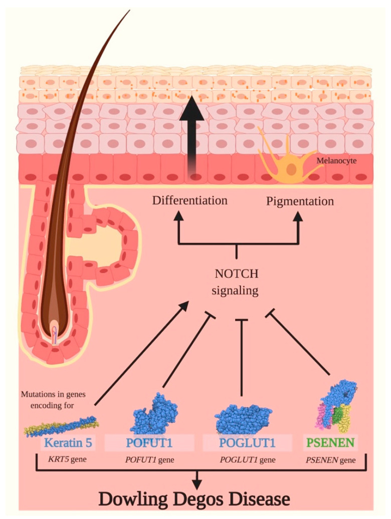
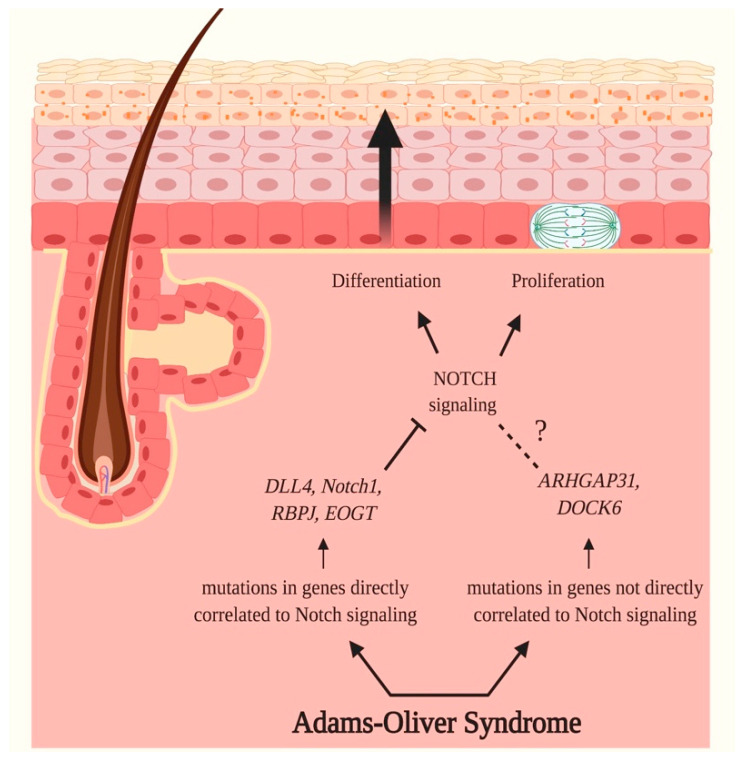
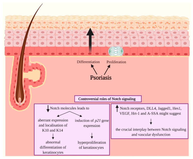
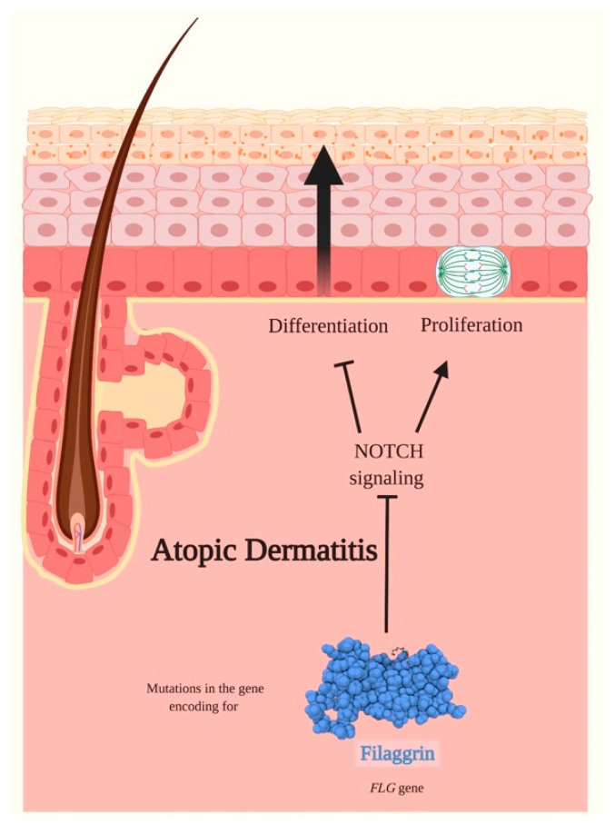
Notch signaling orchestrates the regulation of cell proliferation, differentiation, migration and apoptosis of epidermal cells by strictly interacting with other cellular pathways. Any disruption of Notch signaling, either due to direct mutations or to an aberrant regulation of genes involved in the signaling route, might lead to both hyper- or hypo-activation of Notch signaling molecules and of target genes, ultimately inducing the onset of skin diseases. The mechanisms through which Notch contributes to the pathogenesis of skin diseases are multiple and still not fully understood. So far, Notch signaling alterations have been reported for five human skin diseases, suggesting the involvement of Notch in their pathogenesis: Hidradenitis Suppurativa, Dowling Degos Disease, Adams-Oliver Syndrome, Psoriasis and Atopic Dermatitis. In this review, we aim at describing the role of Notch signaling in the skin, particularly focusing on the principal consequences associated with its alterations in these five human skin diseases, in order to reorganize the current knowledge and to identify potential cellular mechanisms in common between these pathologies.

Keywords: Notch pathway; differentiation; proliferation; skin disorder.


Conflict of interest statement

The authors declare no conflict of interest. The funders had no role in the design of the study; in the collection, analyses, or interpretation of data; in the writing of the manuscript, or in the decision to publish the results.

Figures

Figure 1
Figure 1
Structural organization of human Notch receptors and human canonical Notch ligands. (A) Mammalian cells express four different Notch receptors, Notch1–4. All receptors are single-pass type I transmembrane proteins. The notch extracellular domain (NECD) comprises 29 up to 36 epidermal growth-like factor tandem repeats (EGF-like repeats), followed by the negative regulatory region (NRR), composed by three cysteine-rich Lin12-Notch repeats (LNR-A, -B, -C) and a heterodimerization domain (HD). The single transmembrane domain (TMD) is directly followed by the Notch intracellular domain (NICD) consisting of a Recombination Signal Binding Protein for Immunoglobulin Kappa J Region (RBPjκ) association module (RAM), seven ankyrin repeats (ANK), two nuclear localization signals (NLS) and a transactivation domain (TAD) that retains conserved proline/glutamic acid/serine/threonine-rich motifs (PEST). (B) Five canonical Notch ligands have been described in mammals and they are generally referred to as canonical Delta/Serrate/Lag-2 (DSL) ligands. These proteins belong to the Serrate family of ligands (Jagged1 and Jagged2) and to the Delta-like family of ligands (DLL1, DLL3 and DLL4). The extracellular region possesses a conserved structural organization with an N-terminal domain (NTD), followed by a Delta/Serrate/Lag-2 (DSL) domain flanked by the Delta and OSM-11-like region (DOS) and multiple epidermal-growth-factor-like tandem repeats (EGF-like repeats). The NTD, together with the DSL, DOS and EGF-like motifs, is required for ligand binding to Notch receptors. Jagged1 and Jagged2 possess a juxtamembrane cysteine-rich domain (CRD) not present in Delta-like ligands. In the intracellular region, most Serrate ligands present numerous lysine residues involved in ligand signaling. Always within the intracellular C-terminal region, most DSL ligands express a PSD-95/Dlg/ZO-1 (PDZ) motif that is required for interactions with the cytoskeleton.
Figure 2
Figure 2
Overview of canonical Notch signaling. Notch receptors are newly translated within the endoplasmic reticulum, processed by a furin convertase (S1 cleavage) and subjected to O-linked and N-linked glycosylations in the Golgi compartment. Once on the cell surface, the receptor is activated by binding to a ligand on a juxtaposed cell. Following ligand binding, Notch signaling is initiated when the trans-endocytosis of ligand-receptor complexes in the neighboring cell induces a conformational change in the receptors, leading to the exposure of S2, site cleaved by ADAM metalloproteases (S2 cleavage) to generate a membrane-tethered partition of Notch, namely Notch extracellular truncation (NEXT). The cleavage in S2 exposes the S3 and S4, allowing the subsequent proteolytic cleavage in these sites by the γ-secretase complex (S3/S4 cleavage), ultimately leading to the release of an intracellular active fragment, the NICD. The NICD migrates in the nucleus and binds to CBF1/RBP-Jκ/Su(H)/Lag-1 (CSL), known as RBP-Jκ in vertebrates, that under basal conditions is known to function as a transcriptional repressor by associating with ubiquitous co-repressor (Co-R) proteins, and to transcriptional activators of the Mastermind-like (MAML) family, generating the ternary Notch transcription complex that initiates the transcription of downstream target genes. Membrane trafficking and endocytosis is crucial to regulate the availability of receptors and ligands on the cell surface. In the signal receiving cell, pointed arrows (→) define the orderly succession of events ranging from the synthesis and processing of Notch receptor, to its activation on the plasma membrane and subsequent induction of the transcriptional machinery. In the signal sending cell, the → the trans endocytosis of the ligand-receptor complex, while the rounded arrow (↺) describes the endocytosis of the unbound ligand.
Figure 3
Figure 3
Schematic representation of the skin. The skin is composed of two layers: the epidermis and the dermis. The epidermis, the uppermost partition, is defined as a stratified squamous epithelium comprising the interfollicular epidermis (IFE) and various skin appendages (hair follicles, sebaceous glands and sweat glands). The IFE is primarily composed of progressively differentiated keratinocytes organized in specific layers, which include the basal layer (the deepest portion of the epidermis), stratum spinosum, stratum granulosum and stratum corneum (the most superficial portion of the epidermis). The basal layer comprises mitotically active cells that generate during every cell division process, either stem cells that self-renew or transient amplifying cells that gradually undergo terminal differentiation by migrating upwards towards the stratum corneum. This process requires specific signals released by the various skin appendages and contemplates the acquisition of layer-specific characteristics by keratinocytes, including the expression of epidermal keratins and transcriptional activators. Once in the stratum corneum, keratinocytes are completely keratinized, metabolically inactive and are released through desquamation. The epidermis is physically and functionally separated from the surrounding dermis by the basement membrane. The dermis is a connective tissue layer interposed between the epidermis and the subcutaneous tissue, involved in the protection and support of the skin and the deeper layers, in aiding sensation and in assisting thermoregulation.
Figure 4
Figure 4
Distribution of Notch ligands and receptors in human skin. Notch1–4 receptors and their ligands are involved in the maintenance of epidermal homeostasis principally by regulating proliferation and differentiation programs within the epidermal cells, mediating the balance between proliferating basal progenitors and the terminally derived differentiated progeny, ultimately leading to the formation of the epidermal barrier. The distinctive expression patterns of Notch receptors and their ligands within the different epidermal layers seem to be associated with the activation of layer-specific target genes. Notch1–4 receptors have been reported to be expressed in the suprabasal layers, while Notch1 receptor seems to be widely expressed in the basal layer. Jagged1, Jagged2 and Delta-like 1 (DLL1) are the Notch ligands detected in the epidermis. The expression of Jagged1 has been reported to be predominant in the suprabasal layer, while Jagged2 and DLL1 have been primarily identified in the basal layer.
Figure 5
Figure 5
Hidradenitis Suppurativa and Notch signaling. Hidradenitis Suppurativa (HS) is a chronic inflammatory skin disease affecting the pilosebaceous unit, in which up to 35% of cases present a positive family history. These patients frequently carry mutations in NCSTN, PSEN1 and PSENEN genes, respectively encoding for nicastrin (NCSTN), presenilin 1 (PSEN) and presenilin enhancer 2 (PSENEN) (encoding for protein). These genes encode for three of the four subunits of the γ-secretase multiprotein complex, a transmembrane protease involved in the cleavage of Notch receptors, and their haploinsufficiency results in the dysfunction of the complex. As a consequence, the γ-secretase is not able to cleave and activate Notch receptors and therefore induces an impairment of Notch signaling, ultimately resulting in augmented levels of epidermal cell proliferation and differentiation. Therefore, alterations in Notch signaling might underlie, at least partially, the initial steps of HS onset, which includes infundibular hyperkeratosis and hyperplasia of follicular epithelium that anticipate the follicular occlusion and disruption. Pointed arrows (→) define the induction of the indicated process or associated function, while the truncated arrow (Τ) designates the inhibition of the described process.
Figure 6
Figure 6
Dowling Degos Diseases and Notch signaling. Dowling Degos Disease (DDD) is a rare skin genodermatosis characterized by hyperkeratotic papules and reticulate hyperpigmentation. Classical DDD is induced by mutations in KRT5 gene encoding for keratin 5, and the latter is a fundamental protein required for the synthesis of intermediate filaments, principally in the epidermal cells of the stratum basale. The loss of expression of KRT5 during epidermal differentiation seems to be linked to an activation of Notch signaling, therefore negatively impacting epithelial remodeling. Other genes involved in DDD are POFUT1, POGLUT1 and PSENEN, encoding for GDP-fucose protein O-fucosyltransferase 1 (POFUT1), Protein O-glucosyltransferase 1 (POGLUT1) and presenilin enhancer protein 2 (PSENEN), respectively. POFUT1 mutations lead to nonsense-mediated decay of the transcript, to impaired Notch signaling and to an abnormal pigmentation of the skin. Mutations in POGLUT1 gene presumably cause a nonsense-mediated decay of mRNA resulting in haploinsufficiency, in a blockade of the Notch pathway and in promoting cell differentiation. Pathogenic PSENEN variants impair Notch signaling and cause an aberrant differentiation and pigmentation of the epidermis, leading to reticulate pigmentation, comedones, follicular hyperkeratosis, nodules and scars. Pointed arrows (→) define the induction of the indicated process or associated function, while the truncated arrow (T) designates the inhibition of the described process.
Figure 7
Figure 7
Adams–Oliver Syndrome and Notch signaling. Adams–Oliver Syndrome (AOS) is a rare inherited disorder characterized by aplasia cutis congenita of the scalp, terminal transverse limb defects, vascular anomalies, pulmonary and portal hypertension, and retinal hypervascularization. 71% of identified mutations in AOS cases have been identified in genes directly correlated to Notch signaling, therefore suggesting the crucial role of this pathway in the onset of AOS. Mutations have been registered in Notch1, DLL4, RBP-Jκ and EOGT genes, encoding, respectively, for Notch1, Delta-like 4 ligand (DLL4), Recombination Signal Binding Protein for Immunoglobulin Kappa J Region (RBP-Jκ) and EGF domain-specific O-linked N-acetylglucosamine transferase (EOGT). The identified genetic variants impact the maturation of receptors, receptor-ligand binding and interaction, and ultimately the transcription of target genes. As a consequence, these mutations cause a disruption of Notch signaling, therefore negatively impacting proliferation and differentiation of epidermal cells. The remaining 29% of AOS cases carry causative variants in ARHGAP31, encoding for the Rho GTPase-activating protein 31 (ARHGAP31), and DOCK6, encoding for guanine nucleotide exchange factor (GEF), which are not directly associated with Notch signaling. The products of these genes are regulatory proteins implied in the formation of actin cytoskeleton and are consequently involved in cell morphology, cell migration, survival and division; therefore, variants in these genes are thought to negatively regulate these fundamental processes. Pointed arrows (→) define the induction of the indicated process or associated function, while the truncated arrow (T) designates the inhibition of the described process. The dotted line (---) indicates a proposed association that still needs to be clarified.
Figure 8
Figure 8
Psoriasis and Notch signaling. Psoriasis is defined as a chronic immune-mediated and inflammatory skin disease characterized by hyperproliferation and aberrant differentiation of epidermal cells, primary cutaneous manifestation and a strong genetic predisposition. The role of Notch signaling in Psoriasis seems to be controversial since both downregulations and upregulations of Notch molecules might be responsible for the onset of the skin disease. A decrement in Notch molecules results primarily in an abnormal differentiation of keratinocytes due to an aberrant expression and localization of keratin 10 (K10) and keratin 14 (K14) in the epidermis, and in the induction of p21 gene expression leading to hyperproliferation of keratinocytes. A hyperactivation of Notch signaling has also been registered in the skin of psoriatic patients. High expression of Notch receptors, Jagged1, hairy/enhancer of split 1 (Hes1), vascular endothelial growth factor (VEGF), Ring-box protein HRT1 (Hrt-1) and Acute-phase Serum Amyloid A (A-SSA) have been registered, and they suggest the tight link between Notch signaling and vascular dysfunction in Psoriasis. Pointed arrows (→) define the induction of the indicated process or associated function. The arrows facing upwards (↑) indicate an upregulation, while the arrows facing downwards (↓) designate a downregulation of Notch molecules.
Figure 9
Figure 9
Atopic Dermatitis and Notch signaling. Atopic Dermatitis (AD) is a common, chronic and pruritic inflammatory disease. The pathogenesis of AD is multifactorial and occurs through a combination of disturbed epidermal differentiation with abnormalities in the skin barrier functions, immune dysregulation, and genetic and environmental factors. Mutations in the FLG gene encoding for filaggrin have been registered to be the most common variants in AD familial forms. Filaggrin is a late epidermal differentiation protein that possesses a high keratin-binding affinity, interacts exclusively with intermediate filaments to form highly insoluble keratin scaffolds necessary to guarantee the progressive differentiation of keratinocytes and ultimately form the skin barrier. Loss-of-function mutations in the FLG gene are common in AD and cause a loss of filaggrin expression and therefore to a disturbed epidermal differentiation. Notch signaling is required for the late-stage granular layer differentiation and correct filaggrin processing in the epidermis. A downregulation of Notch receptors expression has been identified in lesional skin of AD. The inactivation of Notch receptors seems to promote the proliferation of epidermal cells leading, amongst other manifestations, to hyperkeratosis and to impair the correct differentiation program in keratinocytes. Pointed arrows (→) define the induction of the indicated process or associated function, while the truncated arrow (T) designates the inhibition of the described process.
Figure 10
Figure 10
Schematic representation of the interactions between Notch signaling, proliferation and differentiation of keratinocytes in skin diseases. The role of Notch signaling, proliferation and differentiation of keratinocytes in Hidradenitis Suppurativa, Dowling Degos Disease, Adams–Oliver Syndrome, Psoriasis and Atopic Dermatitis. The arrows facing upwards (↑) indicate an upregulation, while the arrows facing downwards (↓) designate a downregulation, of Notch signaling, keratinocyte proliferation and differentiation in the various skin diseases.

References

    1. Axelrod J.D., Matsuno K., Artavanis-Tsakonas S., Perrimon N. Interaction Between Wingless and Notch Signaling Pathways Mediated by Dishevelled. Science. 1996;271:1826–1832. doi: 10.1126/science.271.5257.1826. - DOI - PubMed
    1. Talora C., Campese A.F., Bellavia D., Felli M.P., Vacca A., Gulino A., Screpanti I. Notch signaling and diseases: An evolutionary journey from a simple beginning to complex outcomes. Biochimica et Biophysica Acta (BBA)-Mol. Basis Dis. 2008;1782:489–497. doi: 10.1016/j.bbadis.2008.06.008. - DOI - PubMed
    1. Siebel C., Lendahl U. Notch Signaling in Development, Tissue Homeostasis, and Disease. Physiol. Rev. 2017;97:1235–1294. doi: 10.1152/physrev.00005.2017. - DOI - PubMed
    1. Ayaz F., Osborne B.A. Non-Canonical Notch Signaling in Cancer and Immunity. Front. Oncol. 2014;4 doi: 10.3389/fonc.2014.00345. - DOI - PMC - PubMed
    1. Mi H., Muruganujan A., Ebert D., Huang X., Thomas P.D. PANTHER version 14: More genomes, a new PANTHER GO-slim and improvements in enrichment analysis tools. Nucleic Acids Res. 2019;47:D419–D426. doi: 10.1093/nar/gky1038. - DOI - PMC - PubMed

Substances