Review
. 2020 Jun 16;24(1):353.
doi: 10.1186/s13054-020-03062-7.

The vascular endothelium: the cornerstone of organ dysfunction in severe SARS-CoV-2 infection

Affiliations
Review

The vascular endothelium: the cornerstone of organ dysfunction in severe SARS-CoV-2 infection

Stéphanie Pons et al. Crit Care. .

Abstract

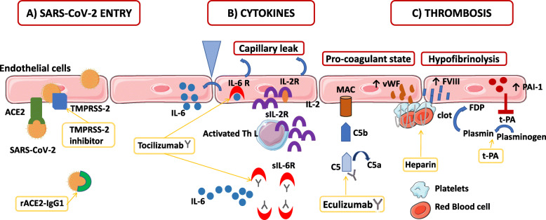
In severe SARS-CoV-2 infections, emerging data including recent histopathological studies have emphasized the crucial role of endothelial cells (ECs) in vascular dysfunction, immunothrombosis, and inflammation.Histopathological studies have evidenced direct viral infection of ECs, endotheliitis with diffuse endothelial inflammation, and micro- and macrovascular thrombosis both in the venous and arterial circulations. Venous thrombotic events, particularly pulmonary embolism, with elevated D-dimer and coagulation activation are highly prevalent in COVID-19 patients. The pro-inflammatory cytokine storm, with elevated levels of interleukin-6 (IL-6), IL-2 receptor, and tumor necrosis factor-α, could also participate in endothelial dysfunction and leukocyte recruitment in the microvasculature. COVID-19-induced endotheliitis may explain the systemic impaired microcirculatory function in different organs in COVID-19 patients. Ongoing trials directly and indirectly target COVID-19-related endothelial dysfunctions: i.e., a virus-cell entry using recombinant angiotensin-converting enzyme 2 (ACE2) and transmembrane protease serine 2 (TMPRSS-2) blockade, coagulation activation, and immunomodulatory therapies, such as anti-IL-6 strategies. Studies focusing on endothelial dysfunction in COVID-19 patients are warranted as to decipher their precise role in severe SARS-CoV-2 infection and organ dysfunction and to identify targets for further interventions.

Keywords: COVID-19; Cytokines; Endothelial cells; Endothelial dysfunction; SARS-CoV-2; Thrombosis.


Conflict of interest statement

SP received a research grant from the French Intensive Care Society, one from the European Society of Intensive Care Medicine, and one from the Zoll foundation. EA has received fees for lectures from MSD, Pfizer, and Alexion. His institution and research group have received support from Baxter, Jazz Pharma, Fisher&Payckle, Gilead, Alexion, and Ablynx. LZ received a research grant from Jazz Pharma.

Figures

Fig. 1
Fig. 1
The role of endothelial cells in SARS-CoV-2 infection and treatment. a Severe acute respiratory syndrome coronavirus 2 (SARS-CoV-2) binds with Angiotensin- converting enzyme 2 (ACE2) on the cell membrane of the host cells. Cell invasion also depends on the presence of the protease Transmembrane protease serine 2 (TMPRSS-2) that is able to cleave the viral spike. The recombinant protein of human ACE2 fused with the Fc region of the human immunoglobulin IgG1 (rACE2-IgG1) binds with high affinity to the receptor-binding domain of SARS-CoV-2. Moreover, a protease TMPRSS2 inhibitor is efficient to block SARS-CoV-2 entry into the endothelial cells (ECs). b In patients diagnosed with severe COVID-19, increased levels of pro-inflammatory cytokines, in particular, the soluble interleukin 2-receptor (IL-2R) and interleukin-6 (IL-6) have been observed. ECs express both IL-6 receptor (IL-6R) and IL-2R on their surface. Soluble IL-2R (sIL-2R) is mostly secreted by activated T helper lymphocytes, but might be also secreted by ECs. Binding of IL-6 and IL-2 on their receptors induces a capillary leak. Moreover, IL-6 signaling induces the secretion by ECs of more IL-6 and other cytokines. Tocilizumab, a humanized anti-IL-6 receptor (IL-6R) antibody that inhibits signal transduction by binding sIL-6R and membrane-bound IL-6R, has emerged as a potential immunomodulatory treatment in COVID-19 patients. c During SARS-CoV-2 infection, endothelial dysfunction and microthrombi formation may be secondary to complement activation and membrane attack complexes deposits on ECs. Eculizumab, a human monoclonal antibody designed to bind to the complement protein C5 with high affinity prevents the generation of the terminal membrane attack complex. EC activation also induces a pro-coagulant state by increasing their production of von Willebrand factor (vWF) and factor VIII (FVIII), which participate in clot formation. Heparin from prophylactic to therapeutic doses has been used in COVID-19 patients to inhibit clot formation. Moreover, during SARS-CoV-2 infection, ECs increase their production of plasminogen activator inhibitor 1 (PAI-1), which inhibits the conversion of plasminogen to plasmin and the degradation of clots inducing a hypofibrinolytic state. Tissue plasminogen activator (t-PA) has been tested in COVID-19 patients for its fibrinolytic effect. ACE2: angiotensin-converting enzyme 2; C5: complement C5; FVIII: factor VIII; FDP: fibrin degradation product; IL-2: interleukin-2; IL-6: interleukin-6; IL-2R: interleukin-2 receptor; IL-6-R: interleukin-6 receptor; MAC: membrane attack complex; PAI 1: plasminogen activator inhibitor 1; rACE2-IgG1: recombinant angiotensin-converting enzyme 2 immunoglobulin G1; SARS-Cov-2: severe acute respiratory syndrome coronavirus 2; sIL-2R: soluble interleukin 2 receptor, sIL-6R: soluble interleukin 6 receptor; Th L: T helper lymphocyte; TMPRSS-2: transmembrane protease serine 2 t-PA: tissue plasminogen activator; vWF: von Willebrand factor

References

    1. Zhu J, Ji P, Pang J, Zhong Z, Li H, He C, et al. Clinical characteristics of 3,062 COVID-19 patients: a meta-analysis. J Med Virol. 2020 [cited 2020 May 5]; Available from: http://doi.wiley.com/10.1002/jmv.25884. - DOI - PMC - PubMed
    1. Marini JJ, Gattinoni L. Management of COVID-19 respiratory distress. JAMA. 2020 [cited 2020 May 5]; Available from: https://jamanetwork.com/journals/jama/fullarticle/2765302. - PubMed
    1. Roumenina LT, Rayes J, Frimat M, Fremeaux-Bacchi V. Endothelial cells: source, barrier, and target of defensive mediators. Immunol Rev. 2016;274:307–329. - PubMed
    1. Daniel AE, van Buul JD. Endothelial junction regulation: a prerequisite for leukocytes crossing the vessel wall. J Innate Immun. 2013;5:324–335. - PMC - PubMed
    1. McKenzie JAG, Ridley AJ. Roles of Rho/ROCK and MLCK in TNF-alpha-induced changes in endothelial morphology and permeability. J Cell Physiol. 2007;213:221–228. - PubMed