. 2020 Oct;10(10):1590-1609.
doi: 10.1158/2159-8290.CD-19-1536. Epub 2020 Jun 16.

Lineage Reversion Drives WNT Independence in Intestinal Cancer

Affiliations

Lineage Reversion Drives WNT Independence in Intestinal Cancer

Teng Han et al. Cancer Discov. 2020 Oct.

Abstract

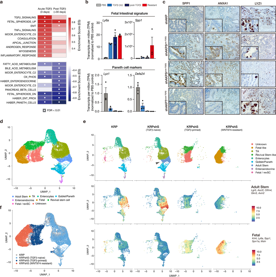
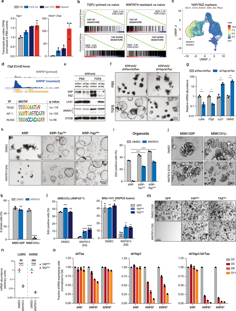
The WNT pathway is a fundamental regulator of intestinal homeostasis, and hyperactivation of WNT signaling is the major oncogenic driver in colorectal cancer. To date, there are no described mechanisms that bypass WNT dependence in intestinal tumors. Here, we show that although WNT suppression blocks tumor growth in most organoid and in vivo colorectal cancer models, the accumulation of colorectal cancer-associated genetic alterations enables drug resistance and WNT-independent growth. In intestinal epithelial cells harboring mutations in KRAS or BRAF, together with disruption of TP53 and SMAD4, transient TGFβ exposure drives YAP/TAZ-dependent transcriptional reprogramming and lineage reversion. Acquisition of embryonic intestinal identity is accompanied by a permanent loss of adult intestinal lineages, and long-term WNT-independent growth. This work identifies genetic and microenvironmental factors that drive WNT inhibitor resistance, defines a new mechanism for WNT-independent colorectal cancer growth, and reveals how integration of associated genetic alterations and extracellular signals can overcome lineage-dependent oncogenic programs. SIGNIFICANCE: Colorectal and intestinal cancers are driven by mutations in the WNT pathway, and drugs aimed at suppressing WNT signaling are in active clinical development. Our study identifies a mechanism of acquired resistance to WNT inhibition and highlights a potential strategy to target those drug-resistant cells.This article is highlighted in the In This Issue feature, p. 1426.


Conflict of interest statement

Conflict of Interest Statement

LED is a scientific advisor for Mirimus Inc.

Figures

Figure 1.
Figure 1.. Engineering of mouse-derived intestinal tumor organoids.
a. Oncoprint derived from MSK IMPACT sequencing and ArcherDx fusion testing, showing co-occurring mutations in WNT, MAPK, TP53 and SMAD4 genes in RSPO fusion-positive human CRCs. b. Schematic representation of the generation of sequentially mutated murine intestinal organoids. c. Representative sanger sequencing chromatogram confirming expected Ptprk-Rspo3 mRNA fusion junction following CRISPR-mediated intrachromosomal inversion. d. Representative Sanger sequencing chromatogram from cDNA, confirming expression of the KrasG12D (G>A) mutant transcript. e. Western blots showing loss of p53 and SMAD4 in independent biological replicates of edited and selected organoids. f. Bright field (upper) and immunofluorescent (lower) images of sequential mutants. Proliferating cells are marked by EdU (Red), and differentiated cells by KRT20 (green).
Figure 2.
Figure 2.. Kras, p53 and Smad4 mutations enable Wnt independence.
a. Bright field and immunofluorescent images of KRP, KRS and KRPS organoids treated with DMSO or WNT974 (upper panel). Bright field and immunofluorescent images of shAKP and shAKPS organoids before and following APC restoration (dox withdrawal) (lower panel). Proliferating cells are marked by EdU (Red), and differentiated cells by KRT20 (green). b. Percentage of EdU-positive cells measured by flow cytometry in KRP, KRS, naive KRPS and resistant KRPS organoids treated with DMSO or WNT974 (n=3 independent biological replicates per condition). During the initial establishment of resistant cells, organoids were treated with PORCN inhibitor continuously for 30 days. After that, resistant cultures were maintained without WNT974. EdU incorporation was assessed following re-treatment with WNT974 for 4 days, at least two weeks after cultures were removed from WNT974. c. qPCR of canonical WNT targets showing WNT signaling is well suppressed in the majority (16/20) of WNT974-resistant quadruple mutant organoid lines and is reactivated in a subset (4/20) of organoid lines. qPCR of canonical WNT targets from WT cells treated with WNT974 is used as a control of maximum WNT pathway suppression. All error bars represent the +/− standard error of the mean (SEM). All statistical tests are two-way ANOVA with Sidak’s correction for multiple comparisons; *p<0.05, **p<0.01, ***p<0.001, ****p<0.0001.
Figure 3.
Figure 3.. TGFβ is required for WNT independence.
a. GSEAs show inflammation-related pathways are enriched in WNT independent lines. b. Western blot analysis showing Smad4 knockdown. KRPshS organoids are generated by transducing KRP organoids with SGEN-shSmad4, then selected by G418 at the concentration of 500ug/mL. c. Bright field images of KRP, KRPshS and KRPT organoids treated with PBS, 5ng/mL TGFβ or 5ng/mL TGFβ plus 10uM TGFBR1 inhibitor LY2157299 as indicated. d. Quantification of TGFβ-induced organoid morphology change. e. Heatmap of TGFβ target genes in KRP and KRPshS acutely treated with TGFβ, or TGFβ plus LY2157299. f. Western blots showing Smad2/3 depletion inhibits the induction of TGFβ downstream targets. g. Bright field images (left) and quantifications (right) showing that Smad2/3 depletion blocks TGFβ-induced spheroid formation. h. Bright field images of KRPshS (pre- and post-TGFβ) and KRPT (post-TGFβ) showing TGFβ priming is required for WNT974 resistance. i. Bright field images of p53 restoration in KRSshP* WNT974-resistant line showing p53 is required for the maintenance of WNT independence. j. EdU flow cytometry on p53 restored WNT974-naive KRSshP* and WNT974-resistant KRSshP* lines. All error bars represent +/− standard error of the mean (SEM). k. Western blot showing the induction of p21 after p53 restoration is inhibited by shCdkn1a in KRSshP*. l. EdU flow cytometry on p53 restored WNT974-naive KRSshP* and WNT974-resistant KRSshP* lines expressing either shRenilla or shCdkn1a. Percentage of relative EdU positive cells is calculated by normalizing the off-Dox EdU percentage to the on-Dox EdU percentage. All error bars represent +/− standard error of the mean (SEM). All statistical tests are two-way ANOVA with Sidak’s correction for multiple comparisons; *p<0.05, **p<0.01, ***p<0.001, ****p<0.0001.
Figure 4.
Figure 4.. The in vivo tumor microenvironment is sufficient to prime cells for WNT independence.
a. Immunohistochemical stains of organoid transplants on dox for 7 weeks (ON DOX), or on dox for 2 weeks then off dox for 5 weeks (OFF DOX). b. Quantification of Ki67 positive epithelial cells in individual tumors from Apc restoration experiments (n≥4 individual tumors). Box and whisker plots represent the full data range (min-max), p-values calculated using two-way ANOVA with Sidak’s correction for multiple comparisons. c. Tumor weight quantification following transplant of different organoid genotypes. Error bars represent +/− standard error of the mean (SEM), n≥4 individual tumors, p-values calculated using a two-sided t-test, with Welsh’s correction. d. Quantification of Ki67 positive epithelial cells in individual tumors from WNT974 treatment experiments (n≥4 individual tumors).
Figure 5.
Figure 5.. TGFβ drives fetal-like lineage reversion.
a. GSEA summary showing pathway enrichment scores for KRPshSmad4 organoids treated for 3 days with TGFβ (acute) and the same organoids one month after TGFβ withdrawal. White dots represent significantly enriched gene sets (FDR<0.01). b. mRNA expression (transcript per million estimates) of fetal intestinal markers (upper) and Paneth cell markers (lower) in KRPshS organoids. Error bars are +/− SEM. c. Immunohistochemical staining of SPP1, ANXA1, and LYZ1 on organoid-derived tumors, as labelled. d. UMAP plot of merged scRNAseq from KRP, KRPshS (pre-TGFβ), KRPshS (post-TGFβ), and KRPshS (WNT974-resistant) organoids, showing identified and putatively identified cell populations. e. UMAP plot (upper) of individual scRNAseq samples shows expansion of Lgr5high adult stem cell population following Smad4 silencing (arrow), and transition of cell lineage following TGFβ-priming and development of WNT-independence. Feature plots (middle and lower) highlight the mean expression of adult stem cell and fetal intestinal markers, as indicated. f. UMAP plot of merged scRNAseq data highlighting specific samples. All error bars represent +/− standard error of the mean (SEM).
Figure 6.
Figure 6.. Yap/Taz is required for lineage reversion and WNT independence.
a. Normalized transcript per million (TPM) shows upregulation of Yap1 and Taz (Wwtr1) transcripts following TGFβ-priming and development of WNT-independence. b. Gene set enrichment analysis (GSEA) plots showing YAP signature is enriched in post-TGFβ KRPshS cells, compared to PBS-treated isogenic organoids, and in WNT974-resistant cells, compared to isogenic WNT974 naïve cells. c. UMAP plot of scRNAseq data showing relative mean gene expression of known YAP/TAZ transcriptional targets. Dotted outline indicates WNT974-resistant population. d. ATACseq profile showing increased accessibility of Ctgf (Ccn2) locus in WNT974-resistant KRPS compared to isogenic WNT974-naive KRPS. Lower panel shows HOMER motif analysis from ATACseq showing top 3 known transcription factor binding sites enriched in WNT974-resistant KRPS organoids compared to WNT974-naive cells. e. Western blot of whole cell lysates from KRPshS/shRen/shRen and KRPshS/shYap/shTaz organoids treated with PBS or 5ng/mL TGFβ as indicated. f. Bright field images of KRPshS/shRen/shRen and KRPshS/shYap/shTaz organoids treated with PBS or 5ng/mL TGFβ as indicated. g. qRT-PCR analysis of gene expression in KRPshS/shRen/shRen and KRPshS/shYap/shTaz organoids treated with 5ng/mL TGFβ. h. Bright field images of KRP, KRP-TAZ4SA and KRP-YAP5SA organoids treated with DMSO or 500nM WNT974 for 4 days. i. Percentage of EdU-positive cells measured by flow cytometry on KRP, KRP-TAZ4SA and KRP-YAP5SA organoids treated with DMSO or 500nM WNT974 for 4 days. j. Bright field images of patient-derived organoid line MSK132P (APC mutant) and MSK121Li (RNF43 mutant) treated with DMSO or 500nM WNT974 for 10 days. k. Percentage of EdU-positive cells measured by flow cytometry on MSK132P and MSK121Li organoids treated with DMSO or 500nM WNT974 for 5 days. l. Percentage of EdU-positive cells measured by flow cytometry on MSK121Li organoids (left) and SNU1411 human RSPO3 fusion CRC cells (right) expressing control vector (GFP) or YAP5SA or TAZ4SA treated with WNT974 in 3D Matrigel culture for 6 days, as indicated. Data represent treatment of 3 independent transductions (n=3, *p<0.05, one-way ANOVA, with Tukey’s correction). m. Bright field images of MSK121Li-GFP, MSK121Li- YAP5SA and MSK121Li-TAZ4SA organoids treated with DMSO or 500nM WNT974 for 30 days. n. qRT-PCR of WNT targets (LGR5 and AXIN2) from WNT-independent MSK121Li organoids treated with DMSO or 500nM WNT974, showing that WNT signaling is not reactivated in these WNT-independent lines. o. Cell competition assays on KRP, KRPS WNT974-naive (KRPSN) and KRPS WNT974-resistant (KRPSR) lines transduced with shTaz, shYap1 and shYap1/shTaz vectors. All error bars represent +/− standard error of the mean (SEM). All statistical tests are two-way ANOVA with Sidak’s correction for multiple comparisons; *p<0.05, **p<0.01, ***p<0.001, ****p<0.0001.

References

    1. Luis TC, Weerkamp F, Naber BA, Baert MR, de Haas EF, Nikolic T, et al. Wnt3a deficiency irreversibly impairs hematopoietic stem cell self-renewal and leads to defects in progenitor cell differentiation. Blood 2009;113(3):546–54 doi 10.1182/blood-2008-06-163774. - DOI - PubMed
    1. Zhao C, Blum J, Chen A, Kwon HY, Jung SH, Cook JM, et al. Loss of beta-catenin impairs the renewal of normal and CML stem cells in vivo. Cancer Cell 2007;12(6):528–41 doi 10.1016/j.ccr.2007.11.003. - DOI - PMC - PubMed
    1. Pinto D, Gregorieff A, Begthel H, Clevers H. Canonical Wnt signals are essential for homeostasis of the intestinal epithelium. Genes Dev 2003;17(14):1709–13 doi 10.1101/gad.267103. - DOI - PMC - PubMed
    1. Fevr T, Robine S, Louvard D, Huelsken J. Wnt/beta-catenin is essential for intestinal homeostasis and maintenance of intestinal stem cells. Mol Cell Biol 2007;27(21):7551–9 doi 10.1128/MCB.01034-07. - DOI - PMC - PubMed
    1. Cancer Genome Atlas N. Comprehensive molecular characterization of human colon and rectal cancer. Nature 2012;487(7407):330–7 doi 10.1038/nature11252. - DOI - PMC - PubMed

Publication types