

Machine learning algorithms reveal unique gene expression profiles in muscle biopsies from patients with different types of myositis
- PMID: 32546599
- PMCID: PMC10461844
- DOI: 10.1136/annrheumdis-2019-216599
Machine learning algorithms reveal unique gene expression profiles in muscle biopsies from patients with different types of myositis
Abstract
Objectives: Myositis is a heterogeneous family of diseases that includes dermatomyositis (DM), antisynthetase syndrome (AS), immune-mediated necrotising myopathy (IMNM), inclusion body myositis (IBM), polymyositis and overlap myositis. Additional subtypes of myositis can be defined by the presence of myositis-specific autoantibodies (MSAs). The purpose of this study was to define unique gene expression profiles in muscle biopsies from patients with MSA-positive DM, AS and IMNM as well as IBM.
Methods: RNA-seq was performed on muscle biopsies from 119 myositis patients with IBM or defined MSAs and 20 controls. Machine learning algorithms were trained on transcriptomic data and recursive feature elimination was used to determine which genes were most useful for classifying muscle biopsies into each type and MSA-defined subtype of myositis.
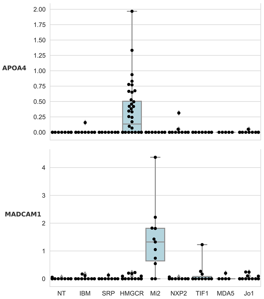
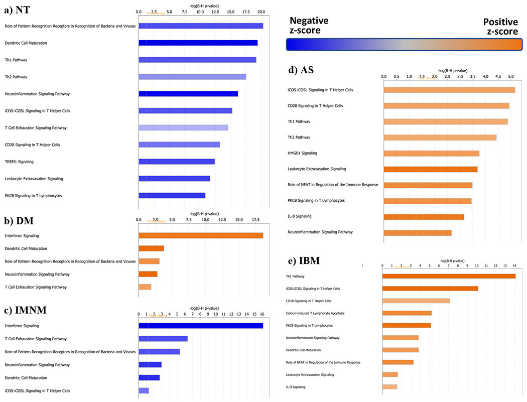
Results: The support vector machine learning algorithm classified the muscle biopsies with >90% accuracy. Recursive feature elimination identified genes that are most useful to the machine learning algorithm and that are only overexpressed in one type of myositis. For example, CAMK1G (calcium/calmodulin-dependent protein kinase IG), EGR4 (early growth response protein 4) and CXCL8 (interleukin 8) are highly expressed in AS but not in DM or other types of myositis. Using the same computational approach, we also identified genes that are uniquely overexpressed in different MSA-defined subtypes. These included apolipoprotein A4 (APOA4), which is only expressed in anti-3-hydroxy-3-methylglutaryl-CoA reductase (HMGCR) myopathy, and MADCAM1 (mucosal vascular addressin cell adhesion molecule 1), which is only expressed in anti-Mi2-positive DM.
Conclusions: Unique gene expression profiles in muscle biopsies from patients with MSA-defined subtypes of myositis and IBM suggest that different pathological mechanisms underly muscle damage in each of these diseases.
Keywords: autoantibodies; autoimmune diseases; autoimmunity; dermatomyositis; polymyositis.
© Author(s) (or their employer(s)) 2020. No commercial re-use. See rights and permissions. Published by BMJ.
Conflict of interest statement
Competing interests: None declared.
Figures

Comment in
-
Gene expression profiles in muscle differ in myositis subtypes.Nat Rev Rheumatol. 2020 Aug;16(8):409. doi: 10.1038/s41584-020-0468-3. Nat Rev Rheumatol. 2020. PMID: 32636494 No abstract available.
-
Correspondence on 'Machine learning algorithms reveal unique gene expression profiles in muscle biopsies from patients with different types of myositis'.Ann Rheum Dis. 2023 Mar;82(3):e61. doi: 10.1136/annrheumdis-2020-219677. Epub 2020 Dec 18. Ann Rheum Dis. 2023. PMID: 33355102 No abstract available.
-
Response to: 'Correspondence on 'Machine learning algorithms reveal unique gene expression profiles in muscle biopsies from patients with different types of myositis'' by Takanashi et al.Ann Rheum Dis. 2023 Mar;82(3):e62. doi: 10.1136/annrheumdis-2020-219767. Epub 2021 Jan 13. Ann Rheum Dis. 2023. PMID: 33441300 Free PMC article. No abstract available.
References
-
- Love LA, Leff RL, Fraser DD, et al. A new approach to the classification of idiopathic inflammatory myopathy: myositis-specific autoantibodies define useful homogeneous patient groups. Medicine; analytical reviews of general medicine, neurology, psychiatry, dermatology, and pediatries 1991;70:360–74. - PubMed
-
- Betteridge Z, McHugh N. Myositis-specific autoantibodies: an important tool to support diagnosis of myositis. Journal of internal medicine 2016;280:8–23. - PubMed
-
- Greenberg SA, Pinkus JL, Pinkus GS, et al. Interferon-alpha/beta-mediated innate immune mechanisms in dermatomyositis. Annals of Neurology 2005;57:664–78. - PubMed
Publication types
MeSH terms
Substances
Supplementary concepts
Grants and funding
LinkOut - more resources
Full Text Sources
Other Literature Sources
Medical
Research Materials
Miscellaneous
