. 2020 May 19:12:3661-3672.
doi: 10.2147/CMAR.S243300. eCollection 2020.

Knockdown of circRAD18 Mitigates Breast Cancer Progression through the Regulation of miR-613/HK2 Axis

Affiliations

Knockdown of circRAD18 Mitigates Breast Cancer Progression through the Regulation of miR-613/HK2 Axis

Hongliang Zang et al. Cancer Manag Res. .

Retraction in

Abstract

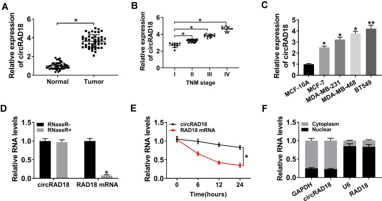
Background: Breast cancer (BC) remains the most prevalent malignancy and the leading cause of cancer death. Circular RNAs (circRNAs) have been discovered to serve as crucial regulators in BC. In the current work, we aimed to study the impact of circRAD18 (hsa_circ_0002453) on BC progression and mechanism governing it.

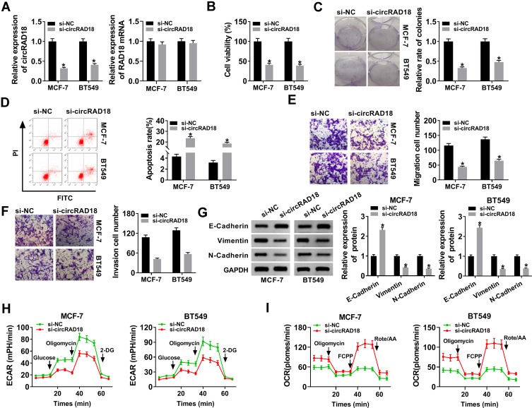
Materials and methods: The expression levels of circRAD18, miR-613 and hexokinase 2 (HK2) mRNA were determined by quantitative real-time polymerase chain reaction (qRT-PCR). CircRAD18 identification was performed using RNase R digestion and actinomycin D assay. Cell viability, colony formation, apoptosis, migration, invasion and glycolysis were measured by Cell Counting Kit-8 assay, colony formation assay, flow cytometry, transwell analysis and extracellular acidification rate detection assay, respectively. Western blot was used to assess the levels of E-Cadherin, Vimentin, N-Cadherin and HK2 protein. The targeted interplay between miR-613 and circRAD18 or HK2 was detected by dual-luciferase reporter assay. Xenograft model assay was performed to observe the role of circRAD18 in vivo.

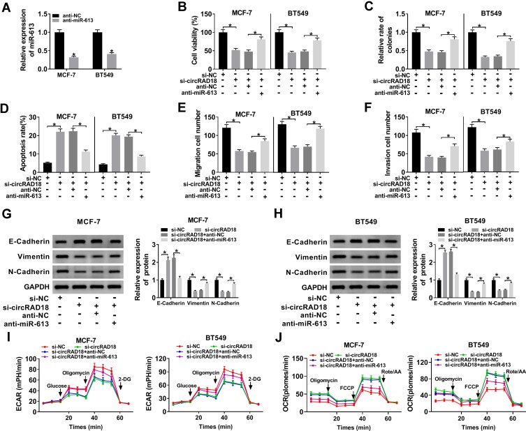
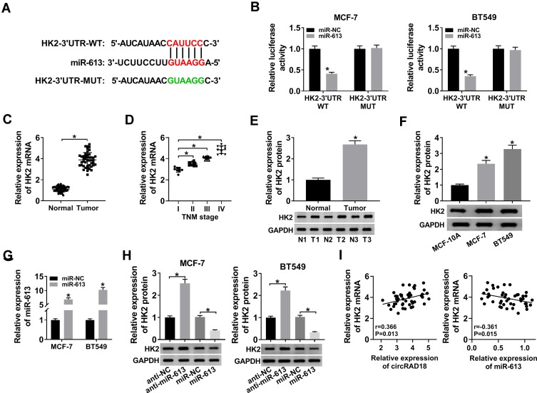
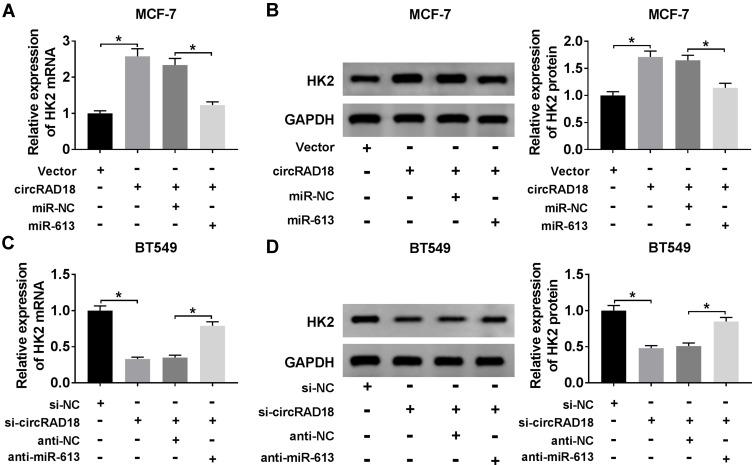
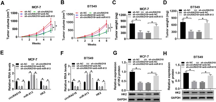
Results: CircRAD18 was highly expressed in BC tissues and cells. CircRAD18 depletion hindered BC cell malignant behaviors, as evidenced by the inhibition in cell viability, colony formation, migration, invasion, epithelial to mesenchymal transition and glycolysis, as well as the promotion in cell apoptosis. CircRAD18 directly interacted with miR-613, and miR-613 mediated the repressive effect of circRAD18 knockdown on BC cell malignant behaviors. Moreover, HK2 was a direct target of miR-613, and circRAD18 positively regulated HK2 expression via sponging miR-613. Additionally, circRAD18 knockdown repressed tumor growth in vivo by miR-613.

Conclusion: Our current work suggested that circRAD18 silencing suppressed BC cell malignant behaviors in vitro and tumor growth in vivo at least partly via the regulation of the miR-613/HK2 axis, highlighting that circRAD18 might be a promising therapeutic target for BC treatment.

Keywords: BC; HK2; circRAD18; malignant behaviors; miR-613.


Conflict of interest statement

The authors declare that they have no financial conflicts of interest.

Figures

Figure 1
Figure 1
Characterization of circRAD18 and it was up-regulated in BC tissues and cells. CircRAD18 expression by qRT-PCR in 45 pairs of BC tissues and matched healthy breast tissues (A), clinical stage (I, II, III and IV) BC tissue samples (B), MCF-10A, MCF-7, MDA-MB-231, MDA-MB-468 and BT549 cells (C). (D) The levels of circRAD18 and linear RAD18 mRNA in BC cells by qRT-PCR after digestion with or without RNase R. (E) The levels of circRAD18 and linear RAD18 mRNA in BC cells after treatment with actinomycin D for the indicated time point. (F) CircRAD18 level in nuclear and cytoplasmic fractions of BC cells by qRT-PCR. *P < 0.05, **P < 0.01.
Figure 2
Figure 2
CircRAD18 silencing retarded the malignant behaviors of BC cells. MCF-7 and BT549 cells were transfected with si-circRAD18 or si-NC. (A) CircRAD18 expression and RAD18 mRNA level by qRT-PCR after 48 h transfection. (B) Cell viability by CCK-8 assay 48 h after transfection. (C) Cell colony formation using a standard colony formation assay after 48 h transfection. (D) Cell apoptosis by flow cytometry after 48 h transfection. (E and F) Cell migration and invasion by transwell assay after 24 h transfection. (G) The levels of E-Cadherin, Vimentin and N-Cadherin by Western blot 48 h after transfection. (H and I) Measurement of ECAR and OCR in transfected cells. *P < 0.05.
Figure 3
Figure 3
CircRAD18 directly interacted with miR-613 in BC cells. (A) Schematic of the complementary site for miR-613 in circRAD18 and the mutated target site. (B) Relative luciferase activity in MCF-7 and BT549 cells cotransfected with circRAD18-WT or circRAD18-MUT and miR-NC mimic or miR-613 mimic. MiR-613 expression by qRT-PCR in 45 pairs of BC tissues and matched healthy breast tissues (C), clinical stage BC (I, II, III and IV) tissue samples (D), MCF-10A, MCF-7, and BT549 cells (E), MCF-7 and BT549 cells transfected with si-NC or si-circRAD18 (F). *P < 0.05.
Figure 4
Figure 4
MiR-613 mediated the repressive impact of circRAD18 deficiency on BC cell malignant behaviors. (A) The expression of miR-613 by qRT-PCR in MCF-7 and BT549 cells transfected with anti-NC or anti-miR-613. MCF-7 and BT549 cells were transfected with si-NC, si-circRAD18, si-circRAD18+anti-NC, or si-circRAD18+anti-miR-613, followed by the determination of cell viability by CCK-8 assay (B), colony formation ability by a colony formation assay (C), cell apoptosis by flow cytometry (D), cell migration (E) and invasion (F) by transwell assay, E-Cadherin, Vimentin and N-Cadherin levels by Western blot (G and H), cell glycolysis by ECAR and OCR measurement (I and J). *P < 0.05.
Figure 5
Figure 5
HK2 was a direct target of miR-613. (A) Schematic of the miR-613-binding sequence in HK2 3ʹ-UTR and the mutant in the target sequence. (B) Relative luciferase activity in MCF-7 and BT549 cells transfected with HK2-3ʹ-UTR-WT or HK2-3ʹ-UTR-MUT and miR-NC mimic or miR-613 mimic. The expression of HK2 in 45 pairs of BC tissues and matched healthy breast tissues (C), clinical stage (I, II, III and IV) BC tissue samples (D), 3 pairs of BC tissues and matched healthy breast tissues (E), MCF-10A, MCF-7, and BT549 cells (F). (G) MiR-613 level by qRT-PCR in MCF-7 and BT549 cells transfected with miR-NC mimic or miR-613 mimic. (H) HK2 protein level by Western blot in MCF-7 and BT549 cells transfected with anti-NC, anti-miR-613, miR-NC mimic, or miR-613 mimic. (I) The correlations between circRAD18, miR-613 and HK2 mRNA expression were determined in BC tissues using the Spearman test. *P < 0.05.
Figure 6
Figure 6
CircRAD18 acted as a sponge of miR-613 to regulate HK2 expression. (A and B) HK2 mRNA expression by qRT-PCR and HK2 protein level by Western blot in MCF-7 cells transfected with Vector, circRAD18, circRAD18+miR-NC mimic, or circRAD18+miR-613 mimic. circRAD18: circRAD18 overexpression vector. (C and D) HK2 mRNA expression by qRT-PCR and HK2 protein level by Western blot in BT549 cells transfected with si-NC, si-circRAD18, si-circRAD18+anti-NC, or si-circRAD18+anti-miR-613. *P < 0.05.
Figure 7
Figure 7
CircRAD18 depletion inhibited tumor growth via regulating miR-613 in vivo. The nude mice were subcutaneously inoculated with sh-circRAD18-transducing MCF-7 or BT549 cells (5.0 × 10⁶). We then performed intratumor injection of PBS, anti-NC or anti-miR-613 one week after implantation. In the end, all mice were sacrificed, and tumor tissues were removed. (A and B) After 7 days implantation, tumor volume measurement was began and implemented every one week. (C and D) Tumor weight was determined. (E and F) The levels of circRAD18, miR-613 and HK2 were assessed by qRT-PCR in xenograft tissues. (G and H) HK2 protein level was detected by Western blot in excised tumor tissues. *P < 0.05.

References

    1. Bray F, Ferlay J, Soerjomataram I, Siegel RL, Torre LA, Jemal A. Global cancer statistics 2018: GLOBOCAN estimates of incidence and mortality worldwide for 36 cancers in 185 countries. CA Cancer J Clin. 2018;68(6):394–424. doi:10.3322/caac.21492 - DOI - PubMed
    1. Collignon J, Lousberg L, Schroeder H, Jerusalem G. Triple-negative breast cancer: treatment challenges and solutions. Breast Cancer (Dove Med Press). 2016;8:93–107. doi:10.2147/BCTT.S69488 - DOI - PMC - PubMed
    1. Qu S, Yang X, Li X, et al. Circular RNA: a new star of noncoding RNAs. Cancer Lett. 2015;365(2):141–148. doi:10.1016/j.canlet.2015.06.003 - DOI - PubMed
    1. Kristensen LS, Hansen TB, Venø MT, Kjems J. Circular RNAs in cancer: opportunities and challenges in the field. Oncogene. 2018;37(5):555–565. doi:10.1038/onc.2017.361 - DOI - PMC - PubMed
    1. Shi P, Sun J, He B, et al. Profiles of differentially expressed circRNAs in esophageal and breast cancer. Cancer Manag Res. 2018;10:2207–2221. doi:10.2147/CMAR.S167863 - DOI - PMC - PubMed

Publication types